eBook - ePub
Radiation Biology of Medical Imaging
- English
- ePUB (mobile friendly)
- Available on iOS & Android
eBook - ePub
Radiation Biology of Medical Imaging
About this book
This book provides a thorough yet concise introduction to quantitative radiobiology and radiation physics, particularly the practical and medical application. Beginning with a discussion of the basic science of radiobiology, the book explains the fast processes that initiate damage in irradiated tissue and the kinetic patterns in which such damage is expressed at the cellular level. The final section is presented in a highly practical handbook style and offers application-based discussions in radiation oncology, fractionated radiotherapy, and protracted radiation among others. The text is also supplemented by a Web site.
Trusted by 375,005 students
Access to over 1.5 million titles for a fair monthly price.
Study more efficiently using our study tools.
Information
CHAPTER 1
Anatomy and Physiology
Keywords
Cell components, homeostasis, tissue growth, tissue repair, organs, organ systems
Topics
- Four main components of a cell
- Four tissue groups
- The difference between tissue growth and tissue repair
- Organs and organ systems
- The role of homeostasis
Introduction
The human body is a complex arrangement of chemicals and chemical reactions. Atoms are combined into specific arrangements creating the chemicals that are used in precise reactions. In addition to orderly reactions, the chemicals combine to form the complex substances that make living cells. Chemicals are nonliving components that allow cells, the basic units of all life, to perform all aspects of life. These characteristics include organization, growth, and reproduction. As can be seen in Fig. 1.1, the organization and structure of the body begins with chemicals and progresses through greater levels of organization, beginning simply with cells and ending with the entire human body.
Figure 1.1 Organization of the body, beginning with chemicals combining into simple atoms and progressing through cells, tissues, organs, and, finally, the whole body. From Tortora and Nielsen (2012), figure 1.1, p. 5.
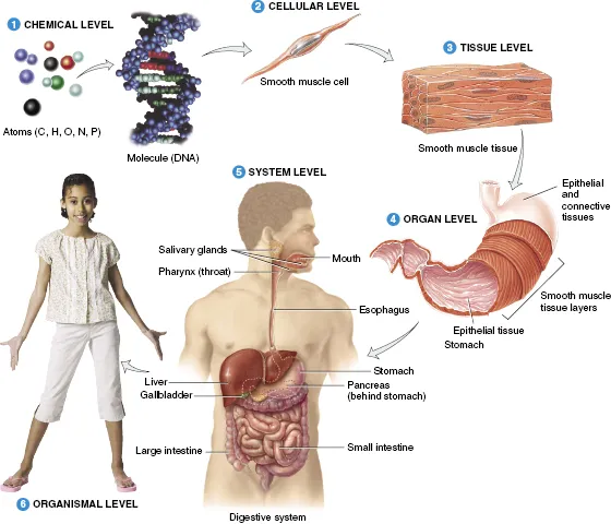
The cell is the simplest structure of the human body. As the levels of organization expand, so does the complexity of the system. Groups of cells with the same, or similar, functions gather to form tissues. For instance, the primary function of pancreatic cells is to produce insulin whereas cells of the kidney aid in the filtration of blood. When a group of similar tissues function together, they become known as an organ. Most organs have several roles and belong to multiple organ systems. An organ system consists of multiple organs that function together and benefit the body as a whole. For example, the respiratory system, which consists primarily of the lungs, allows carbon dioxide to be exchanged for oxygen in the blood. The blood then delivers oxygen to cells throughout the body.
In this chapter, the levels of organization in the human body will be discussed: beginning with the cell, moving through tissue and organ function, and ending with homeostasis.
Mammalian Cell Components
Cells are the smallest viable component of all living organisms. Organisms can be either unicellular, containing only one cell, or multicellular, containing many cells. The human body is multicellular and made up of approximately 100 trillion, 1014, cells. These cells are split into over 200 different types within the body. Each cell type is responsible for a specific function, but, despite differences in structure and function, there are four basic components contained in every cell. These features are the cell membrane, cytoplasm, cellular organelles, and genetic material.
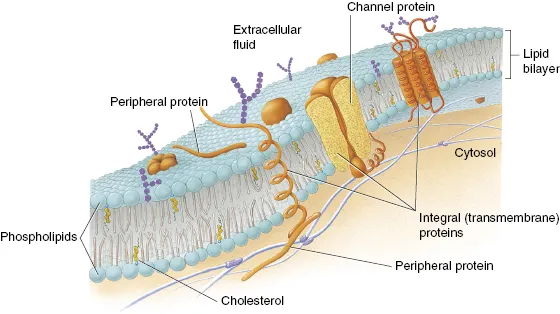
The cell membrane, or plasma membrane, is responsible for the separation of the internal environment of the cell from the external environment. The membrane is primarily constructed of phospholipids, which form a bilayer that makes most of the membrane. Phospholipids allow for movement of lipid-soluble substances into and out of the cell by simple diffusion through the membrane itself. In addition to phospholipids, cholesterol is interspersed throughout the membrane. Cholesterol strengthens the structure of the membrane by decreasing its fluidity. Another vital component of the cell membrane is protein. Protein molecules, like cholesterol, are embedded in the membrane. These proteins have several different functions within the membrane. The functions include forming protein channels and acting as transporters and as receptor sites. Protein channels permit passage of molecules, such as water or other ions, into the cell unabated. Transport proteins, or carrier enzymes, also assist with the movement of molecules into or out of the cell. Receptor proteins are primarily located on the outer side of the membrane. The receptors are used to transmit signals into the cell from external signals. These signals include the absorption of hormones or signaling chemicals. Although the plasma membrane is the outer boundary of the cell, it is not a static, wall-like structure. Shown in Fig. 1.2 is the basic structure of a plasma membrane.
Figure 1.2 Current concept of the structure of the plasma membrane. Cholesterol is interspersed sporadically in one side of the phosopholipid bilayer, whereas proteins more commonly span both phosopholipid layers. From Tortora and Nielsen (2012), figure 2.2, p. 30.
In addition to the cell membrane, each cell is filled with cytoplasm. Cytoplasm is an aqueous substance that resides between the outer cell membrane and the nucleus. The cytoplasm is made of water, salts, and organic molecules and accounts for 70% of the cell volume. Many of the chemical reactions that occur within the cell, such as glycolysis, occur within the cytoplasm. Other cell processes, such as cell division, are also contained within the cytoplasm.
Although organelles are contained within the cytoplasm, they are separated into their own class of cellular components. Organelles are specialized subunits within the cell. Each organelle performs a specific function within the cell. For instance, ribosomes are responsible for transcribing DNA, a vital function for protein synthesis and cell survival. Others, like the mitochondria, are responsible for producing energy. An appropriate analogy of an organelle is that of an organ within the body. Each organ is confined within the body and performs a specific function. In a similar manner, each organelle, confined within the cell, performs a particular function to help maintain the life of the cell. Most organelles are encompassed by individual membranes. These membranes, similar to the outer cell membrane, allow flow of material into and out of the organelle. A typical animal cell, with associated organelles, is shown in Fig. 1.3. Keep in mind that Fig. 1.3 is for a typical mammalian cell. Red blood cells in the human body do not contain organelles. This enables them to deliver a greater amount of oxygen to the body.
Figure 1.3 Diagram of a typical animal cell showing selected organelles and general organization within the cellular membrane. From Tortora and Nielsen (2012), figure 2.1, p. 29.
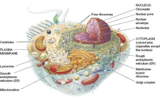
Another component of mammalian cells is genetic material. Genetic material, more commonly known as DNA, is located within the nucleus of each mammalian cell. Similar to other organelles, the nucleus is surrounded by a separate membrane, called the nuclear envelope. This membrane regulates the passage of substances into and out of the nucleus. It also localizes and protects the DNA within the cell. DNA is responsible for encoding messages for everything from the development of physical characteristics, such as hair and eye color, to when a cell should proliferate. The nucleus is the largest of the intercellular organelles and is often referred to as the control center of the cell. Although the nucleus controls the function of mammalian cells, red blood cells do not contain a nucleus. Since red blood cells do not divide once mature, there is no need for maintaining DNA and, hence, no need for a nucleus.
Tissue Groups
Cells are organized into groups that have similar structure and function. Once these cells have gathered together, they become known as tissue. Individual tissues are arranged into characteristic patterns of cells that are specialized for particular functions. The human body consists of four main tissue groups. These tissue classifications are epithelial, connective, muscle, and nervous. The following descriptions of each tissue group will explain basic differences in structure and function.
Epithelial tissue is the covering or lining found on many body surfaces. If the epithelial tissue is a cover, it is located primarily on the outside of the body. The skin is the cover that assists in keeping the inside of the body safe from environmental hazards. When epithelial tissue is utilized as a lining, it is located inside the body. The respiratory system is lined with epithelial tissue that aids in the protection of the lungs. Within the primary classification of the epithelium, the cells are divided into three additional groups that differentiate between cell shapes and functions. These are squamous, cuboidal, and columnar.
Each of the three types of e...
Table of contents
- Cover
- Title page
- Copyright page
- Acknowledgments
- Introduction
- CHAPTER 1: Anatomy and Physiology
- CHAPTER 2: The Cell
- CHAPTER 3: Radiation Characteristics and Units
- CHAPTER 4: Radiation Interactions with Tissue
- CHAPTER 5: Cell Survival Curves
- CHAPTER 6: DNA and Genetics
- CHAPTER 7: Radiation Damage and Repair of Cells
- CHAPTER 8: Normal and Malignant Cells
- CHAPTER 9: Radiation Effects on Tissues and Organs
- CHAPTER 10: Whole Body Radiation Effects
- CHAPTER 11: Radiation Treatment of Cancer
- CHAPTER 12: Radiation Biology of Diagnostic Imaging
- CHAPTER 13: Nuclear Medicine Radiation Biology
- CHAPTER 14: Environmental Radiation
- CHAPTER 15: Regulations and Risk
- CHAPTER 16: Biological Effects of Ultrasound
- CHAPTER 17: Biological Effects of Magnetic Resonance Imaging
- Answers to Odd-Numbered Questions
- Index
Frequently asked questions
Yes, you can cancel anytime from the Subscription tab in your account settings on the Perlego website. Your subscription will stay active until the end of your current billing period. Learn how to cancel your subscription
No, books cannot be downloaded as external files, such as PDFs, for use outside of Perlego. However, you can download books within the Perlego app for offline reading on mobile or tablet. Learn how to download books offline
Perlego offers two plans: Essential and Complete
- Essential is ideal for learners and professionals who enjoy exploring a wide range of subjects. Access the Essential Library with 800,000+ trusted titles and best-sellers across business, personal growth, and the humanities. Includes unlimited reading time and Standard Read Aloud voice.
- Complete: Perfect for advanced learners and researchers needing full, unrestricted access. Unlock 1.5M+ books across hundreds of subjects, including academic and specialized titles. The Complete Plan also includes advanced features like Premium Read Aloud and Research Assistant.
We are an online textbook subscription service, where you can get access to an entire online library for less than the price of a single book per month. With over 1.5 million books across 990+ topics, we’ve got you covered! Learn about our mission
Look out for the read-aloud symbol on your next book to see if you can listen to it. The read-aloud tool reads text aloud for you, highlighting the text as it is being read. You can pause it, speed it up and slow it down. Learn more about Read Aloud
Yes! You can use the Perlego app on both iOS and Android devices to read anytime, anywhere — even offline. Perfect for commutes or when you’re on the go.
Please note we cannot support devices running on iOS 13 and Android 7 or earlier. Learn more about using the app
Please note we cannot support devices running on iOS 13 and Android 7 or earlier. Learn more about using the app
Yes, you can access Radiation Biology of Medical Imaging by Charles A. Kelsey,Philip H. Heintz,Gregory D. Chambers,Daniel J. Sandoval,Natalie L. Adolphi,Kimberly S. Paffett in PDF and/or ePUB format, as well as other popular books in Medicine & Radiology, Radiotherapy & Nuclear Medicine. We have over 1.5 million books available in our catalogue for you to explore.