![]()
SECTION 1 THE HORSE (Equus caballus)
PLATES
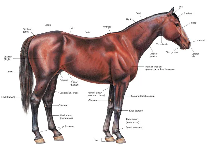
1.1 Right lateral view of a stallion.
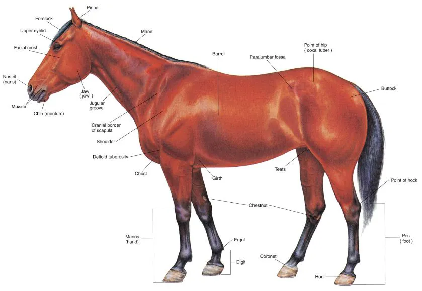
1.2 Left lateral view of a mare.
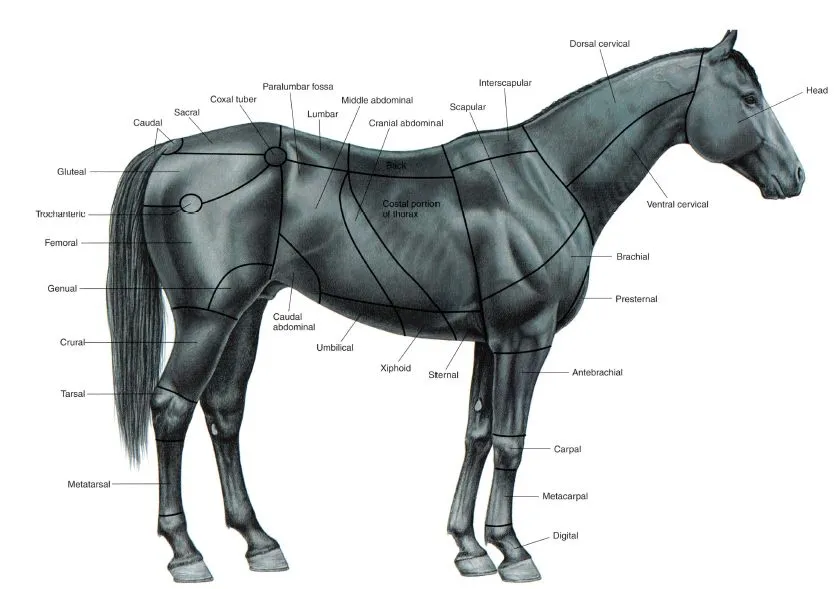
1.3 Body regions of the horse.
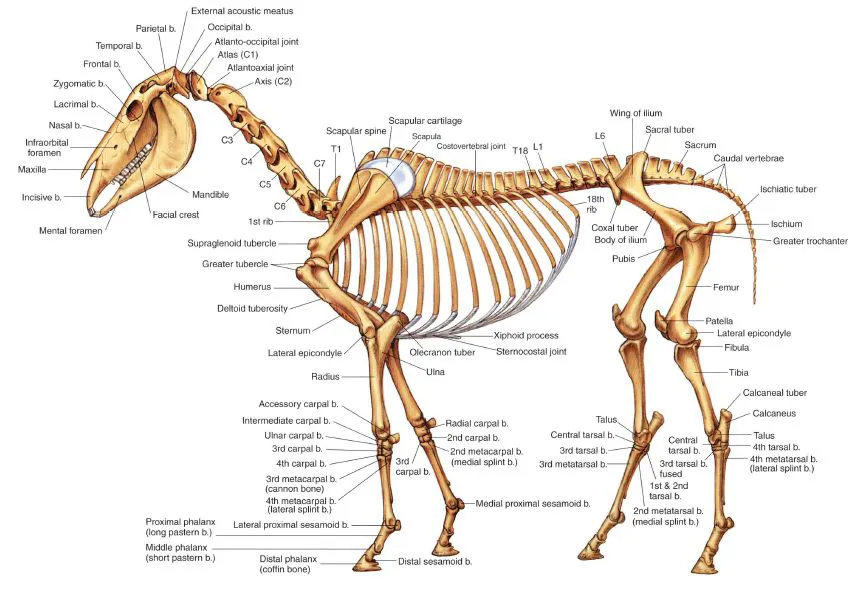
1.4 Skeleton of the horse.
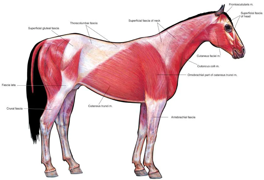
1.5 Cutaneous muscles and major fasciae of the stallion.
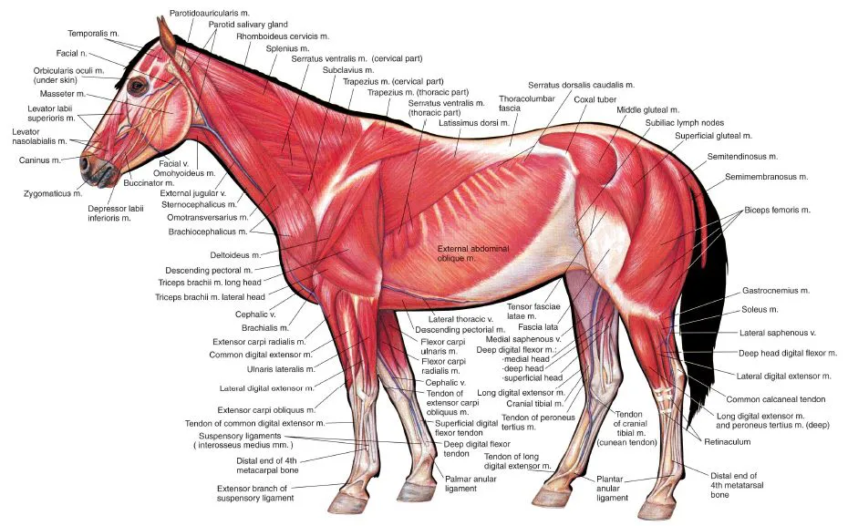
1.6 Superficial muscles and veins of the mare.
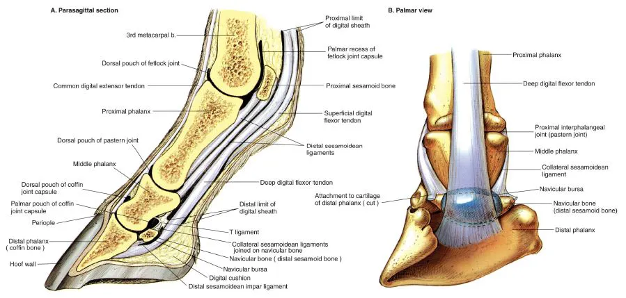
1.7 A. Parasagittal section of the equine digit. B. Palmar (plantar) view of major structures of the digit.
1.8 Relations of the hoof.
1.9 Stay apparatus of the equine forelimb.
1.10 Stay apparatus and reciprocal apparatus of the hindlimb.
1.11 Deep muscles and in situ viscera of the stallion.
1.12 Deep cervical muscles, major joints, and in situ viscera of the mare.
1.13 Median section of the horse's head.
1.14 A. Occlusal (grinding) surfaces of an equine lower first incisor tooth related to continuous eruption and wear. B. Complete dentition of the male horse circa 5 years of age.
1.15 Isolated stomach and intestines of the horse.
1.16 Equine cecum, large (ascending) colon, and transverse colon in situ.
1.17 Clinical condition: Right dorsal displacement of the large colon.
1.18 Clinical condition: Left dorsal displacement of the large colon.
1.19 Reproductive organs, urinary organs, liver, heart, and adjacent major vessels related to the skeleton of the stallion.
1.20 Heart and some adjacent major vessels, abdominal and pelvic viscera, and udder (mammary glands) of the mare.
1.21 Relations of the reproductive organs of the stallion.
1.22 Relations of the reproductive organs of the mare.
1.23 Neonatal organs of the foal.
1.24 Major arteries of the mare.
1.25 Major veins of the stallion. Portal system excluded.
1.26 Lymph nodes and vessels of the horse.
1.27 Central and somatic nervous system of the stallion.
1.28 Autonomic nervous system of the mare.
PLATE 1.1 Right lateral view of a stallion.
PLATE 1.2 Left lateral view of a mare.
PLATE 1.3 Body regions of the horse. Right lateral view.
PLATE 1.4 Skeleton of the horse. Left lateral view. C = cervical vertebra, T = thoracic vertebra, L = lumbar vertebra, b = bone
PLATE 1.5 Cutaneous muscles and major fasciae of the stallion. Right lateral view, m = muscle
PLATE 1.6 Superficial muscles and veins of the mare. Left lateral view. m = muscle, n = nerve, v = vein
PLATE 1.7 A. Parasagittal section of the equine digit. B. Palmar (plantar) view of major structures of the equine digit. Navicular bursa obscures joining of collateral sesamoidean ligaments on the navicular bone, b = bone
PLATE 1.8 Relations of the hoof. A. Separation of the hoof to show its relations to regions of the coriuni. B. Three-dimensional dissection to show relations of the hoof wall, coronary and laminar corium, and distal phalanx. C. Solar surface of...