Learning features include: carefully selected labeling helps students learn and remember structures and relationships; male and female of species are depicted on facing pages so topographic anatomy can be compared; structures common to various animals are labeled several times, whereas unique structures are labeled on one or two species so students can make rapid distinctions of the structures peculiar to certain animals; and an introduction that provides readers with a background in nomenclature and anatomic orientation so they can benefit from the atlas even if they lack training in anatomy.
The Atlas depicts topographic relationships of major organs in a simple, yet technically accurate presentation that's free from extraneous material so that those using the atlas can concentrate on the essential aspects of anatomy. It will be an invaluable resource for veterinary students, teachers and practitioners alike.
eBook - ePub
Color Atlas of Small Animal Anatomy
The Essentials
- English
- ePUB (mobile friendly)
- Available on iOS & Android
eBook - ePub
Color Atlas of Small Animal Anatomy
The Essentials
About this book
This new resource provides a basic foundation in small animal anatomy for students of veterinary medicine, animal science, and veterinary technology. Extraordinary accuracy and beautiful original artwork make this a truly unique learning tool that includes the anatomy of all organ systems in the dog, cat, rabbit, rat, and guinea pig - all described in a consistent manner.
Trusted by 375,005 students
Access to over 1.5 million titles for a fair monthly price.
Study more efficiently using our study tools.
Information
SECTION 1 THE DOG
PLATES
1.1 Surface view of the dog (Beagle)
1.2 Surface view of the bitch (Chesapeake Bay Retriever)
1.3 Body regions
1.4 Skeleton
1.5 Cutaneous muscles and major fasciae of the dog
1.6 Superficial muscles of the bitch
1.7 Deep muscles of the dog
1.8 Deep cervical muscles, major joints, and in situ viscera of the bitch
1.9 Paraxial and medial view of the third digit
1.10 Palmar views of the major structures of the forepaw; plantar view of the major structures of the hindpaw
1.11 Median section of the head and dentition
1.12 Eye and adnexal ocular structures
1.13 Nasal fossae
1.14 External, middle and inner ear
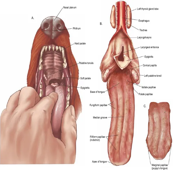
1.15 Oral cavity, tongue, and esophagus
1.16 Ventral view of abdominal structures
1.17 Large intestine, anus and anal sacs
1.18 Body cavities and serous membranes
1.19 Thoracic, abdominal and pelvic viscera related to the skeleton of the dog
1.20 Thoracic, abdominal and pelvic viscera related to the skeleton of the bitch
1.21 Hip joint
1.22 Location of major endocrine organs
1.23 Relations of the reproductive organs of the dog
1.24 Relations of the reproductive organs of the bitch
1.25 Major veins
1.26 Major arteries
1.27 Lymph nodes and vessels
1.28 Central nervous system and spinal nerves
1.29 Autonomic nervous system
1.30 Brain
PLATE 1.1 Right lateral view of a male Beagle dog.
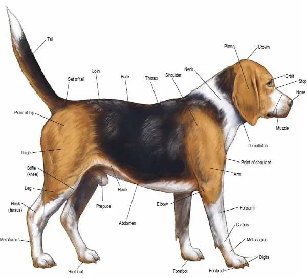
PLATE 1.2 Left lateral view of a female Chesapeake Bay Retriever.
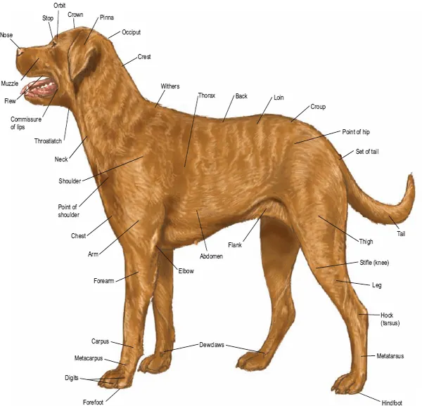
PLATE 1.3 Body regions.
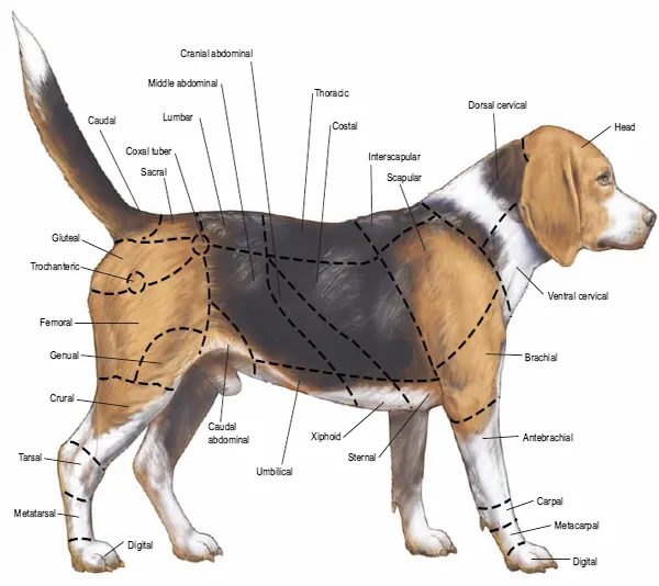
PLATE 1.4 Skeleton; C = Cervical vertebrae, T = Thoracic vertebrae, L = Lumbar vertebrae.
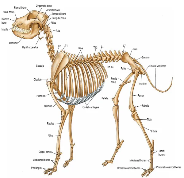
PLATE 1.5 Cutaneous muscles and major fasciae of the dog.
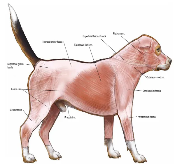
PLATE 1.6 Superficial muscles of the bitch.
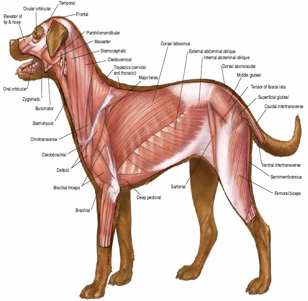
PLATE 1.7 Deep muscles of the dog.
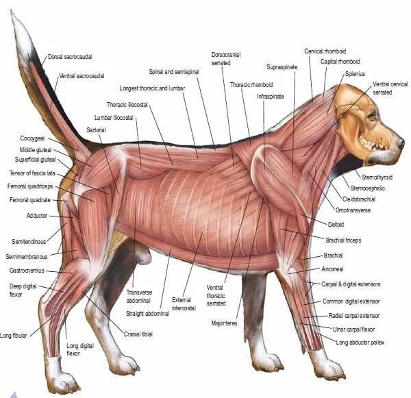
PLATE 1.8 Deep cervical and back muscles (m.), major joints, and in situ viscera of the bitch.
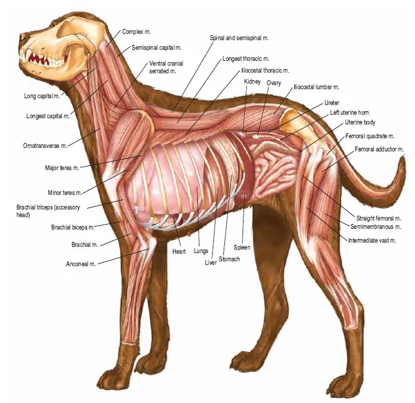
PLATE 1.9 Forepaw: A. Medial superficial view, B. Medial deep view.
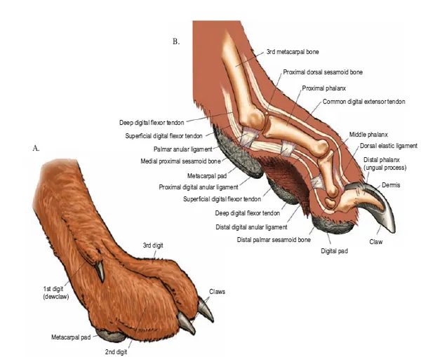
PLATE 1.10 A. Palmar view of the forepaw; B. Palmar view of superficial dissection of forepaw; C. Plantar view of hindpaw; D. Plantar view of superficial dissection of hindpaw.
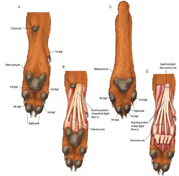
PLATE 1.11 A. Medial sagittal section of head. B. Skull and dentition (M- molar, I- incisor, C - canine, P - premolar).
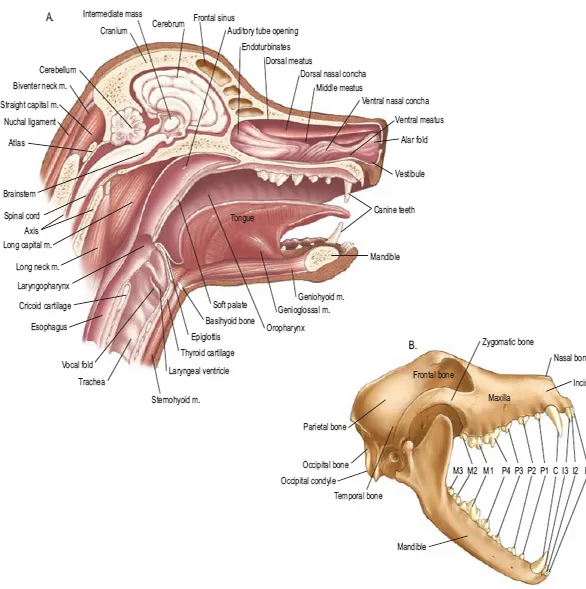
PLATE 1.12 A. The eye and adnexal ocular structures. B. Nasolacrimal duct. C. Median section of eye.
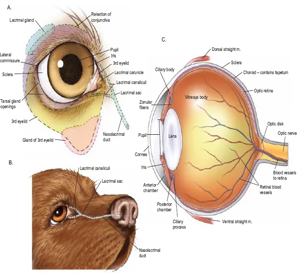
PLATE 1.13 Rt. nasal fossa. A. External nares. B. Nasal cartilages. C. Medial sagittal section of the muzzle.
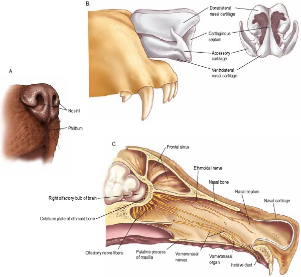
PLATE 1.14 The ear. A. Coronal section of the head and external ear. B. Middle and inner ear.
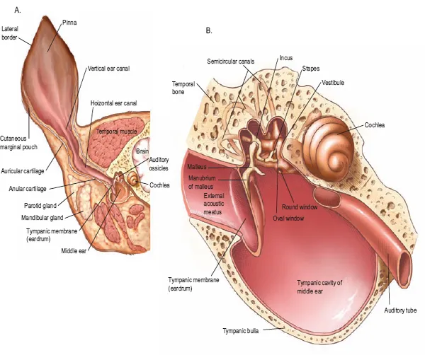
PLATE 1.15 Oral cavity, tongue, and esophagus
P...
Table of contents
- COVER
- CONTENTS
- TITLE PAGE
- COPYRIGHT
- INTRODUCTION
- NOMENCLATURE AND ANATOMIC ORIENTATION
- SECTION 1 THE DOG
- SECTION 2 THE CAT
- SECTION 3 THE RABBIT
- SECTION 4 THE RAT
- SECTION 5 THE GUINEA PIG
- INDEX
Frequently asked questions
Yes, you can cancel anytime from the Subscription tab in your account settings on the Perlego website. Your subscription will stay active until the end of your current billing period. Learn how to cancel your subscription
No, books cannot be downloaded as external files, such as PDFs, for use outside of Perlego. However, you can download books within the Perlego app for offline reading on mobile or tablet. Learn how to download books offline
Perlego offers two plans: Essential and Complete
- Essential is ideal for learners and professionals who enjoy exploring a wide range of subjects. Access the Essential Library with 800,000+ trusted titles and best-sellers across business, personal growth, and the humanities. Includes unlimited reading time and Standard Read Aloud voice.
- Complete: Perfect for advanced learners and researchers needing full, unrestricted access. Unlock 1.5M+ books across hundreds of subjects, including academic and specialized titles. The Complete Plan also includes advanced features like Premium Read Aloud and Research Assistant.
We are an online textbook subscription service, where you can get access to an entire online library for less than the price of a single book per month. With over 1.5 million books across 990+ topics, we’ve got you covered! Learn about our mission
Look out for the read-aloud symbol on your next book to see if you can listen to it. The read-aloud tool reads text aloud for you, highlighting the text as it is being read. You can pause it, speed it up and slow it down. Learn more about Read Aloud
Yes! You can use the Perlego app on both iOS and Android devices to read anytime, anywhere — even offline. Perfect for commutes or when you’re on the go.
Please note we cannot support devices running on iOS 13 and Android 7 or earlier. Learn more about using the app
Please note we cannot support devices running on iOS 13 and Android 7 or earlier. Learn more about using the app
Yes, you can access Color Atlas of Small Animal Anatomy by Thomas O. McCracken,Robert A. Kainer,David Carlson in PDF and/or ePUB format, as well as other popular books in Medicine & Veterinary Medicine. We have over 1.5 million books available in our catalogue for you to explore.