Key concepts have also been added to each chapter to better promote learning, and terms are now defined throughout the text, with the definitions collected into a glossary. User-friendly and well-illustrated with charts, reference values, algorithms and photomicrographs, Hematology Techniques and Concepts for Veterinary Technicians, Second Edition is a key reference for veterinary technicians and veterinary technology students.

eBook - ePub
Hematology Techniques and Concepts for Veterinary Technicians
- English
- ePUB (mobile friendly)
- Available on iOS & Android
eBook - ePub
Hematology Techniques and Concepts for Veterinary Technicians
About this book
Now in full color, Hematology Techniques and Concepts for Veterinary Technicians, Second Edition is a thorough update to this introduction to the fundamental concepts of collecting, handling, and preparing hematology samples. Covering the basics of blood composition, cell morphology, and sample collection, handling, and preparation, the book is designed specifically for veterinary technicians and students to gain a full understanding of why each test is performed and ensure accurate test results. In addition to addressing advances in technology, equipment, and test techniques throughout, a new chapter covers automated testing, and a companion website provides review questions and images from the book for download at www.wiley.com/go/voigt.
Trusted by 375,005 students
Access to over 1.5 million titles for a fair monthly price.
Study more efficiently using our study tools.
Information
Chapter 1
Introduction to the Hematology Laboratory
Key Concepts
In the hematology laboratory, manual and automated test techniques are used in the study of blood and other body cells and fluids.
Blood tests obtained in the laboratory are used by the clinician for screening the health status of an animal or in making a diagnosis of a disease condition.
Quality control of laboratory procedures is essential to ensure that test results reflect the true status of the patient.
Hematology is the study of blood and the tissues that form, store, or circulate blood cells. Examination of the blood is a very common and useful procedure for several reasons. Blood bathes all the other cells of the body carrying nutrients, oxygen, and waste products, and is exposed to almost all metabolic processes of these cells, often reflecting any alteration from normal function. Blood is essential in water and electrolyte balance, temperature control, and the functioning of the immune system, which is the defense mechanism of the body. Obtaining, studying, and testing a blood sample is a relatively easy way of gathering information on many parts of the body.
Even though the term “hematology” literally means “the study of blood,” many of the techniques of collection, sample preparation, and cell identifications learned by the technician can be applied to other regions of the body, such as joint fluid, cerebrospinal fluid, thoracic and abdominal fluids, aspirants from abnormal growths or swellings, and cells collected from mucous membranes (e.g., the oral cavity, trachea, or vagina).
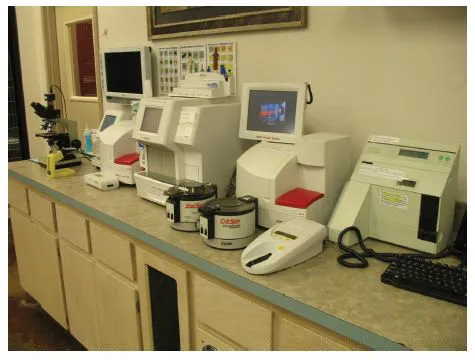
Veterinary technicians should expect to encounter a wide range of laboratory facilities and instrumentation in various veterinary clinics. This can vary from a separate room fully equipped with the latest automated analyzers to a small area on one counter with a microscope, slides, and stains in smaller clinics (Figs. 1.1, 1.2, and 1.3). Even if the clinic sends out all of its hematology tests to a commercial lab, or to the local human hospital, the technician is still usually responsible for correctly collecting, preparing, and mailing or delivering the sample as well as reporting and recording the results.
Hematology is one of several specialties in the field of clinical pathology, a field that encompasses any manual or automated laboratory procedure used on the animal to aid in diagnosing a clinical condition. Clinical chemistry, parasitology, and urinalysis are additional classical aspects of clinical pathology. Other diagnostic tools used by the veterinarian that qualify as clinical pathology under this definition are microbiology, diagnostic imaging (radiology and ultrasound), and the microscopic interpretation of biopsies (tissue samples).
Figure 1.1 Some clinics may have limited laboratory equipment and may routinely send samples to hospital or commercial laboratories.
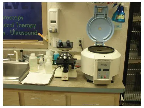
Figure 1.2 Technicians will find clinics with an area dedicated to laboratory work with some of the latest automated equipment.
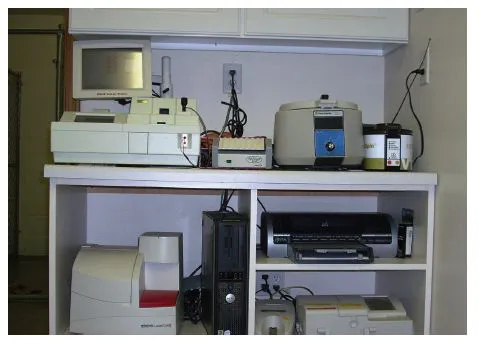
Figure 1.3 Larger veterinary hospitals often have a complete laboratory room that may be staffed by one or more full-time laboratory technicians.
Uses and Benefits of Hematology Results
The most common use of the hematology laboratory is to screen the general health of an animal and to assess its overall ability to transport oxygen and defend against infectious agents. Hematology results provided by the technician are also used by the veterinarian as tools that, when combined with the history, physical examination, and other laboratory findings, help to form a diagnosis. Although occasionally a blood test will yield a definitive diagnosis (e.g., blood parasites), most laboratory results should be viewed as large or small pieces of a diagnostic puzzle that must be assembled.
Hematology results may also indicate a course of treatment for the animal. An example of this is the use of hematology to differentiate between anemia caused by internal hemorrhage and that caused by bone marrow depression. Another example is finding specific infectious organisms within blood cells. Laboratory findings may also suggest other beneficial tests, such as a bone marrow biopsy.
Serial sampling, which is the collection and testing of a series of blood samples over a period of time (hours, days, or even weeks), can demonstrate the severity of a disease process and the ability of the animal to respond. By combining these results with clinical evaluation, the veterinarian will be better able to understand the disease process and make a prognosis for the patient.
It has been said that a clinician who relies entirely on laboratory results to make a diagnosis is probably inexperienced and a clinician who claims not to need a laboratory is uninformed.
Limitations of Laboratory Findings
The validity and usefulness of both manual and automated laboratory results can be influenced by many factors that should be understood by the technician, so they may be eliminated or minimized as much as possible. Technical errors associated with individual tests are discussed in later chapters, but those of general laboratory procedures are addressed in this section.
The first potential problems arise in the collection and handling of the sample. The blood sample, or any other tissue sample, should not be subjected to traumatic physical forces, such as being forced rapidly through a small needle or violently shaken, or to extreme temperatures of heating or freezing. Care must be taken to avoid contaminating the sample with foreign material such as dirt, infectious agents, chemicals, or even water. Test results are always more meaningful with fresh samples, but when this is not possible samples should be refrigerated since blood, like other organic material, begins to degenerate after removal from the body. A more extensive list of sample handling errors is found in Chapter 4.
Quality control, or more accurately “quality assurance,” should be considered a laboratory commitment, rather than a laboratory problem. Whether the test procedures are performed manually, carried out by automated equipment, or even sent to a local hospital or a commercial or state laboratory, it is essential to know that the results reflect the patient’s status and not a difference in machines, technicians, or techniques.
Test results often vary with different test conditions, and the “normal” animal may routinely test higher or lower than published “normal” ranges. Commercial and state laboratories usually provide their established reference values for each test being performed on each species with the laboratory report. Clinical laboratories should establish reference values for any manual or automated test procedure performed in the clinic. For reference values to be statistically valid, a large number of tests must be performed, which may be difficult in a small clinical setting. By keeping a record of all tests run (and data on the patients), plus checking medical records for other similar test results, clinics can establish a database to compare with published values. At a minimum, tests on several known normal or control samples should be run for each procedure by each technician who will be performing the tests. These control samples can also be sent to the outside laboratory being routinely used and the results compared with the in-house results. Some slight variation in results should be expected, but they should fall within the established range for that procedure. Paired samples, half sent to an outside source and half run in-house for comparison, can also be quite instructive. Running such tests will ensure that current test procedures and results are reliable, or the tests can be used to establish reference readings for a new technique, technician, or a piece of equipment. The frequency of checking in-house tests will vary with the type and a number of tests performed and often varies from daily to weekly to monthly but should never be overlooked.
Another area of quality control that must not be overlooked is the variation in skill levels, accuracy, and care of the technician, especially in manual procedures. While the decision about which laboratory test to perform in the clinic or hospital and which to send out depends a great deal on the cost-effectiveness and timely availability of outside services, the number of tests being performed should also be considered. Routinely, if a test is not being done at least a few times per week, it does not provide the laboratory personnel with adequate opportunities to become skilled at, and comfortable with, the procedure or the equipment.
The veterinary technician is often responsible for collecting, preparing, and examining the sample and reporting the results. All counts or measurements should be done on a “blind” basis, that is, with no comparisons to normals or expectations of changes that could lead to bias in reported results. Since the technician may be the only person to actually see the sample, it is imperative that all observations, whether normal or abnormal, be recorded. Often the “comments” section of a lab report is as informative as the recorded results.
Laboratory Safety
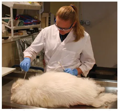
Although most diseases of domestic animals routinely handled by the technician are not communicable to humans (zoonotic), the ones that tend to be quite severe, possibly fatal. All biological samples, whether blood or other body tissues and fluids, should be handled as if potentially infectious. Routine hand washing and disinfection of glassware and working areas is essential. There should be no drinking, eating, use of tobacco products, or other hand-to-mouth activities, nor any storage of food or beverages in the laboratory area. A laboratory coat should be worn at all times, long hair confined (especially around Bunsen burners and centrifuges!), and sandals, open-toed shoes, or canvas shoes should be avoided. Laboratory gloves should be worn whenever potentially infectious samples are being handled (Fig. 1.4). All disposable pointed or cutting instruments (e.g., needles and scalpel blades) should be placed in an appropriate “sharps” container, such as an empty gallon jug or commercial container, prior to disposal. Laboratory safety policies should not only be posted in the laboratory area but also be read and followed.
Figure 1.4 A laboratory smock should be worn at all times, and protective gloves, eyewear, and confining of long hair are important when working around potentially infectious or hazardous conditions.
Review Questions
1. What is the major function of the hematology laboratory?
2. Compare the benefits and limitations of the hematology laboratory.
3. What is meant by the term “quality control” in relation to reporting test results?
4. Why is laboratory safety important to the hematology technician?
Chapter 2
Composition of Blood
Key Concepts
Blood is a tissue composed of a fluid called plasma and seven cellular elements called erythrocytes, neutrophils, eosinophils, basophils, lymphocytes, monocytes, and platelets.
The cellular elements of the blood can be identified by their individual characteristics and the staining qualities of their cellular contents on a prepared glass slide.
Blood is a type of connective tissue, and collecting a blood sample is essentially taking a tissue biopsy. Blood is composed of cells surrounded by a noncellular substance, just like other connective tissues, such as fibrous tissue, bone, or cartilage. The major difference is, of course, that the extracellular substance in blood is liquid, called plasma. This characteristic, plus the fact that much of this “tissue” is located near the surface of the animal, makes collecting a sample of blood comparatively easier than sampling deeper, more solid organs and tissues.
PCV, Buffy Coat, and Plasma
If drawn blood is kept from clotting by the addition of an anticoagulant (see Chapter 4), we can separate the blood into its various components. If allowed to set undisturbed (or placed in a centrifuge to speed up the rate of separation of the components), three distinct layers will appear: the heavy red cells on the bottom, the lighter white cells in the middle, and the plasma on the top (Fig. 2.1).
The bottom one-third to one-half of the tube will be dark red and will contain the heaviest (most dense) structures, the erythrocytes (red blood cells). The measurement of the percentage of these cells in the blood is called the packed cell volume (PCV) or hematocrit (Hct) and is commonly one of the first blood evaluations performed (see Chapter 6).
Directly on the top of the red blood cells will be a relatively thin, white to tan layer called the buffy coat. This layer contains the leukocytes (white blood cells) and the platelets. The thickness of this layer can be a basis for a rough estimate of the relative numbers of these cells in the sample. The occasional appearance of a light pink to red color in the buffy coat indicates the entrapment of some lighter density erythrocytes in this layer.
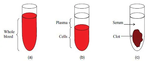
At the top of the tube will be the fluid portion of the blood sample, called plasma since the blood has been separated without clotting. If the blood had been allowed to clot, several proteins in the fluid would have been used to form the clot and the fluid would then be called serum (Fig. 2.2). The plasma color will vary from clear to straw to yellow-orange (due to species variation, diet, and physiologic or pathologic conditions) but should be transparent. The technician should always observe the visual characteristics of the plasma, especially any cloudiness, abnormal coloration, or layering, and write descriptive comments on the laboratory report. (Remember, the technician may be the only one to see the sample and should pass on the information!)
Figure 2.1 The components of unclotted blood.
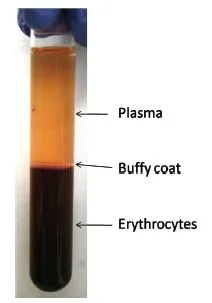
Figure 2.2 Physical difference between plasma and serum. (a) Mixed fluid and cells in whole blood. (b) With anticoagulant added, cells separate from plasma. (c) Without an anticoagulant, cells and clotting factors separate from serum.
Blood Cells
In order to see and evaluate individual blood cells, a drop of blood must be spread on a slide and stained. This is one of the most common and important hematology procedures. Techniques for making and staining the blood smear, as well as suggested methods of viewing the slide under the microscope to ensure optimum examination of the cells, are given in Chapters 5 and 6.
There are seven “formed elements” (cells or cell fragments) found in the blood:
| Erythrocytes (red blood cells or RBCs) | (1) |
| Leukocytes (white blood cells or WBCs) | |
| Granulocytes | |
| Neutrophils | (2) |
| Eosinophils (acidophils) | (3) |
| Basophils | (4) |
| Agranulocytes | |
| Lymphocytes | (5) |
| Monocytes | (6) |
| Thrombocytes (platelets) | (7) |
A diagram of the basic differences in appearance of each of these cells is shown in Fig. 2.3. A brief description of the appearance and function of each is then presented to allow the reader to become familiar with and begin to identify these cells. More detailed descriptions are given in later chapters.
Figure 2.3 The seven “formed elements” (cells and cell fragments) ...
Table of contents
- Cover
- Title page
- copyright
- Preface
- Acknowledgments
- Chapter 1: Introduction to the Hematology Laboratory
- Chapter 2: Composition of Blood
- Chapter 3: Blood Volume and Effects of Blood Loss
- Chapter 4: Blood Collection and Handling
- Chapter 5: Blood Smears and Staining
- Chapter 6: Routine Hematology Laboratory Tests
- Chapter 7: Automated Laboratory Methods and Instruments
- Chapter 8: Leukocyte Cell Types and Functions
- Chapter 9: Introduction to the Immune System
- Chapter 10: Erythrocyte Form, Function, and Indices
- Chapter 11: Erythrocyte Abnormalities
- Chapter 12: Anemias and Polycythemias
- Chapter 13: Hemostasis and Coagulation
- Chapter 14: Hematopoiesis and Bone Marrow Examination
- Chapter 15: Collection and Handling of Cytology Samples
- Glossary
- Recommended Reading
- Index
Frequently asked questions
Yes, you can cancel anytime from the Subscription tab in your account settings on the Perlego website. Your subscription will stay active until the end of your current billing period. Learn how to cancel your subscription
No, books cannot be downloaded as external files, such as PDFs, for use outside of Perlego. However, you can download books within the Perlego app for offline reading on mobile or tablet. Learn how to download books offline
Perlego offers two plans: Essential and Complete
- Essential is ideal for learners and professionals who enjoy exploring a wide range of subjects. Access the Essential Library with 800,000+ trusted titles and best-sellers across business, personal growth, and the humanities. Includes unlimited reading time and Standard Read Aloud voice.
- Complete: Perfect for advanced learners and researchers needing full, unrestricted access. Unlock 1.5M+ books across hundreds of subjects, including academic and specialized titles. The Complete Plan also includes advanced features like Premium Read Aloud and Research Assistant.
We are an online textbook subscription service, where you can get access to an entire online library for less than the price of a single book per month. With over 1.5 million books across 990+ topics, we’ve got you covered! Learn about our mission
Look out for the read-aloud symbol on your next book to see if you can listen to it. The read-aloud tool reads text aloud for you, highlighting the text as it is being read. You can pause it, speed it up and slow it down. Learn more about Read Aloud
Yes! You can use the Perlego app on both iOS and Android devices to read anytime, anywhere — even offline. Perfect for commutes or when you’re on the go.
Please note we cannot support devices running on iOS 13 and Android 7 or earlier. Learn more about using the app
Please note we cannot support devices running on iOS 13 and Android 7 or earlier. Learn more about using the app
Yes, you can access Hematology Techniques and Concepts for Veterinary Technicians by Gregg L. Voigt,Shannon L. Swist in PDF and/or ePUB format, as well as other popular books in Medicine & Veterinary Medicine. We have over 1.5 million books available in our catalogue for you to explore.