![]()
p.1
1 Organization and Function of the Nervous System
Our behaviors, including our thoughts, sensations, emotions, memories, and even our states of consciousness, are all a result of complex interactions between neurons distributed throughout our brains. These neurons form elaborate systems that communicate their activity by releasing small amounts of transmitter substances which act both on receiving neurons as well as on the neuron sending the message. In order for us to understand just how drugs act to treat certain psychological conditions, we must first understand the intricate and sometimes subtle ways in which neurons function to regulate our behaviors. We must also appreciate the complex systems of neurons within the brain that specialize in different functions including movement, emotions, learning and memory, and our motivational states.
The average human brain weighs approximately 1,400 grams (or roughly three pounds) and contains nearly 200 billion neurons. Each of these neurons may in turn communicate with just a few or as many as tens of thousands of other neurons. How the structure and organization of neurons and their surrounding environment allows for such communication will be the topic of the first part of this chapter. We will then describe the structures and functions of systems within the brain that allow humans and other organisms to function in, and adapt to, their continuously changing environments. This background will be necessary for us to understand how psychological disorders may arise and just how drugs might help to alleviate them.
The Structure and Function of Neurons
As mentioned above, the brain contains approximately 200 billion individual nerve cells or neurons. These neurons are the basic units of the brain as well as the rest of the nervous system. Neurons vary in shape, size, and other characteristics according to their location and their specific function.
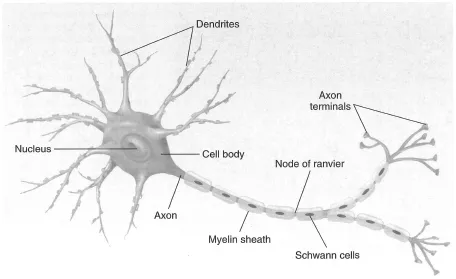
There are three major classes of neurons: sensory neurons, motor neurons, and interneurons. Sensory or afferent neurons carry ascending messages to the central nervous system (CNS) from receptors in the skin, ears, nose, eyes, as well as some organs, muscles, and joints. The brain and sometimes the spinal cord interpret these messages and send appropriate responses through descending motor or efferent neurons, which lead to sensory organs, muscles, glands, and other peripheral tissues to control movement and the functioning of glands, sensory organs, and other tissues. Interneurons reside only within the CNS and function to bridge communication between sensory and motor neurons. Without these connecting neurons, sensory messages would never result in the appropriate bodily response. Interneurons also communicate with each other throughout the nervous system. Although neurons vary in size, shape, and function, they share four common structures: the cell body, the dendrites, the axon, and the terminal buttons (see Figure 1.1).
p.2
Cell Body or Soma
The cell body or soma is the largest part of the neuron. It contains structures that control the cell’s metabolic functions (cell respiration and metabolism). It also contains the nucleus which contains the cell’s genetic information encoded in DNA. The membrane of the cell body can have receptors and receive messages from other neurons, although the cell body is not typically the cell’s primary receiving target.
Dendrite
Neurons typically receive messages from other cells at a collection of extensions from the cell body called dendrites, which branch out from the cell body like roots of a tree. (The word dendrite comes from the Greek word for tree.) Dendrites may receive information from a few to thousands of surrounding neurons. The more extensive the neuron’s network of dendrites, the more connections can be made with other neurons. Interneurons in the brain typically contain far more dendritic branches than neurons in the spinal cord or the peripheral nervous system. Signals received by dendrites are passed on to the membrane of the cell body where excitatory and inhibitory signals are integrated and a decision is made whether to transmit the signal along its axon.
p.3
Axon
The axon is typically an extended branch of the cell that functions to transmit the electrical signal from the surface of the cell body towards receiving cells. The point on the cell body where both the axon and the electrical signal originate is called the axon hillock. The electrical signal is transmitted along the entire length of the axon, which may range from several feet in length in spinal cord and peripheral nervous system (PNS) neurons to fractions of millimeters in neurons within the brain. The axon may divide into two or more major branches called collaterals, thereby increasing its capacity to communicate with other neurons. Axons may be myelinated or unmyelinated. Myelin is a type of glial cell that wraps around the axon providing it with insulation. Most peripheral axons are myelinated, and most (but not all) of the axons in the brain are unmyelinated. Myelin serves both to insulate the axon, much like insulation on a wire, and to increase the speed of conduction along the axon. It is myelin that gives brain tissue, which is normally grayish brown, a white color (white vs. gray matter).
Terminal Button
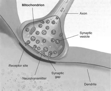
The transmitting end of the axon consists of small bulblike structures known as terminal buttons (see Figure 1.2). The terminal buttons store and release neurotransmitters which either excite or inhibit adjacent neurons. Terminal buttons are also where neurotransmitter substances are taken back into the cell after their release. The structure that allows for neurotransmitter reuptake is a protein called a reuptake transporter. These transporter proteins will be given considerable attention throughout this text as they are the site where many psychotropic drugs are designed to work.
p.4
Once the recycled neurotransmitters, or their precursor chemicals, have been taken back into the terminal button, it is further transported back into synaptic vesicles where it is stored for subsequent release. The amount of neurotransmitter available in synaptic vesicles for release depends on the availability of its metabolic precursors, on the frequency of cell firing, and whether or not it is receiving messages to turn down neurotransmitter synthesis and release at specialized receptors on the terminal button.
Neural Transmission
In order for a message to travel from neuron to neuron, it must move from the terminal button at the end of one neuron’s axon to the dendrites or cell body of an adjacent neuron. The process by which impulses are transmitted in the CNS is called neural transmission and it involves both electrical and chemical processes.
Within the PNS, messages are transmitted along the extended axonal fibers of both motor and sensory neurons that are contained within bundles of neural fibers called nerves. The multitudes of neural circuits or pathways within the CNS are made up of perhaps hundreds of thousands of individual neurons. These fibers extend as continuous structures from sensory receptors or muscles to the CNS. For example, a sensory message from a pain receptor in the skin of your finger is transmitted along a single axonal fiber that extends the length of your arm to a point at which it enters the spinal cord and transfers its message to an interneuron.
Neuron Electrical Activity
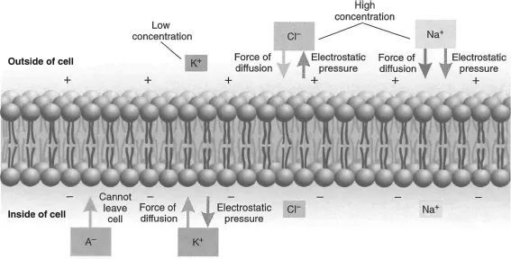
All cells, including neurons, are enclosed in a lipid membrane composed of two layers of lipid molecules called a lipid bilayer (see Figure 1.3). This membrane acts as a kind of skin that permits the cell to maintain an internal environment different from the fluid outside of the membrane. The membrane communicates with its external environment through specialized integrated proteins that are distributed throughout the lipid structure. These proteins function to carry glucose to internal cell structures and to carry metabolic waste back out. They also serve to carry chemical ions back and forth across the membrane. These ions carry either a positive or a negative electrical charge and therefore change the membrane’s electrical potential. Ions that are particularly important in neural transmission are negatively charged organic ions (An–), chlorine ions (Cl–), positively charged sodium ions (Na+), and potassium ions (K+). If the cell membrane did not act as a barrier, these ions would be equally distributed both inside and outside of the neuron. However, the negative organic ions do not pass through the cell membrane to the surrounding fluid and the membrane is only semi permeable to other ions. For instance, sodium and chlorine ions pass through only when gates are open for them. These gates, called ion channels, are actually proteins embedded in the cell’s membrane and they become activated by changes in the membrane potential or by the presence of specific chemicals on their surface.
p.5
Resting Potentials
There are essentially two forces acting on these charged ions. The first force is diffusion, which is the pressure on ions to distribute themselves equally in a medium. That is, to move from high to lower concentrations. Perfume diffuses from an open bottle throughout a room. The second force is electrostatic. Ions of similar charge repel each other as do similarly charged sides of a magnet. This electrostatic force acts to move ions towards the opposite charge and away from a similar charge. When these two forces are at equilibrium the neuron is said to be in its resting state. The distribution of negatively and positively charged ions on either side of the membrane determines the cell’s electrical potential during this resting state. This resting potential is therefore mostly determined by the concentrations of charged ions in the fluids on both sides of the cell membrane. The ion transport proteins that are embedded in the cell membrane can also contribute to the resting potential to some extent because they also carry an electric charge.
The negative and positive charges are unequal on either side of the membrane when the two forces are in equilibrium, so its interior has a negative electrical potential with respect to its exterior. This phenomenon is due primarily to the negatively charged organic ions on the inside and a high concentration of positively charged sodium ions outside the membrane. Most neurons at rest (that is, when their membrane potential is not changing) have a net negative charge of about −70 millivolts (70/1,000 of a volt) relative to their outside environment. The membrane is said to be in a polarized state when the neuron is at rest (see Figure 1.4).
This differential charge gives the resting neuron a state of potential energy known as the resting potential. In ...