Case-based and heavily illustrated, Intraoperative and Interventional Echocardiography: Atlas of Transesophageal Imaging, 2nd Edition covers virtually every clinical scenario in which you're likely to use TEE. Drs. Donald C. Oxorn and Catherine M. Otto provide practical, how-to guidance on transesophageal echocardiography, including new approaches and state-of-the-art technologies. More than 1, 500 images sharpen your image acquisition and analysis skills and help you master this challenging technique.- Real-world cases and abundant, detailed figures and tables show you exactly how to proceed, step by step, and get the best results.- Each case begins with a brief presentation and discussion, and integrates clinical echocardiography, surgical pathology, and other imaging data.- Clear descriptions of preoperative pathology guide you in choosing the best approach to common problems.- The practice-based learning approach with expert commentary for each case helps you retain complex information and apply it in your daily practice.- Every chapter has been thoroughly revised, with discussions of new technology and new techniques, including several techniques that are on the verge of becoming mainstream.- New chapters cover current transcatheter valve therapies and device closures.- Expert Consult eBook version included with purchase. This enhanced eBook experience allows you to search all of the text, figures, images, and references from the book on a variety of devices.
eBook - ePub
Intraoperative and Interventional Echocardiography
Atlas of Transesophageal Imaging E-Book
- 448 pages
- English
- ePUB (mobile friendly)
- Available on iOS & Android
eBook - ePub
Intraoperative and Interventional Echocardiography
Atlas of Transesophageal Imaging E-Book
About this book
Trusted by 375,005 students
Access to over 1.5 million titles for a fair monthly price.
Study more efficiently using our study tools.
Information
Chapter 1
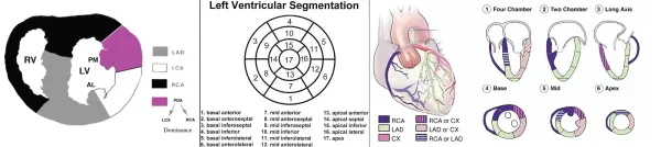
Coronary artery disease
Visualization of the coronary arteries and regional wall motion
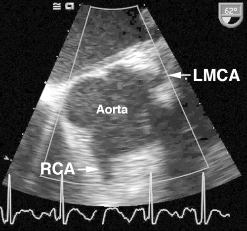
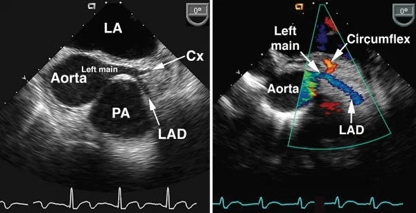
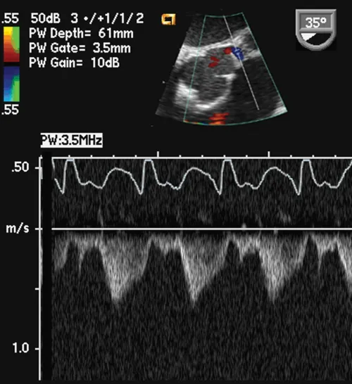
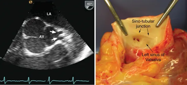
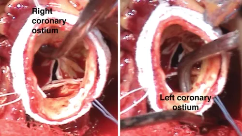
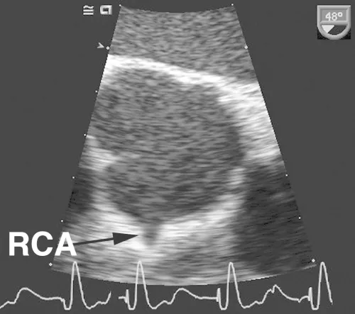
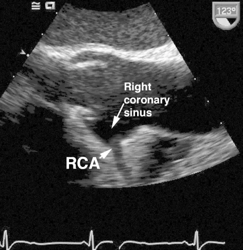
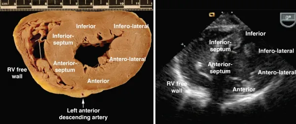
CASE 1-1 Normal coronary arteries
Comments
As shown in these examples, the proximal coronary arteries can often be visualized on TEE. The left main coronary artery arises from the left coronary sinus of Valsalva, is easily visualized in over 85% of patients and has a normal diameter of 4.2 ± 0.7 mm, with a slightly smaller average diameter in women (3.5 mm) compared with men (4.3 mm). The left main coronary artery bifurcates into the left anterior descendin...
Table of contents
- Cover image
- Title page
- Table of Contents
- Copyright
- Foreword
- Preface
- Acknowledgments
- Abbreviations
- 1. Coronary artery disease
- 2. Mitral valve disease
- 3. Aortic valve disease
- 4. Endocarditis
- 5. Surgical prosthetic valves
- 6. Right-sided valve disease
- 7. Adult congenital heart disease
- 8. Hypertrophic cardiomyopathy
- 9. Pericardial disease
- 10. Diseases of the great vessels
- 11. Masses
- 12. Mechanical circulatory support
- 13. Transcatheter valve therapies
- 14. Transcatheter closure devices
- Index
Frequently asked questions
Yes, you can cancel anytime from the Subscription tab in your account settings on the Perlego website. Your subscription will stay active until the end of your current billing period. Learn how to cancel your subscription
No, books cannot be downloaded as external files, such as PDFs, for use outside of Perlego. However, you can download books within the Perlego app for offline reading on mobile or tablet. Learn how to download books offline
Perlego offers two plans: Essential and Complete
- Essential is ideal for learners and professionals who enjoy exploring a wide range of subjects. Access the Essential Library with 800,000+ trusted titles and best-sellers across business, personal growth, and the humanities. Includes unlimited reading time and Standard Read Aloud voice.
- Complete: Perfect for advanced learners and researchers needing full, unrestricted access. Unlock 1.5M+ books across hundreds of subjects, including academic and specialized titles. The Complete Plan also includes advanced features like Premium Read Aloud and Research Assistant.
We are an online textbook subscription service, where you can get access to an entire online library for less than the price of a single book per month. With over 1.5 million books across 990+ topics, we’ve got you covered! Learn about our mission
Look out for the read-aloud symbol on your next book to see if you can listen to it. The read-aloud tool reads text aloud for you, highlighting the text as it is being read. You can pause it, speed it up and slow it down. Learn more about Read Aloud
Yes! You can use the Perlego app on both iOS and Android devices to read anytime, anywhere — even offline. Perfect for commutes or when you’re on the go.
Please note we cannot support devices running on iOS 13 and Android 7 or earlier. Learn more about using the app
Please note we cannot support devices running on iOS 13 and Android 7 or earlier. Learn more about using the app
Yes, you can access Intraoperative and Interventional Echocardiography by Donald Oxorn,Catherine M. Otto in PDF and/or ePUB format, as well as other popular books in Medicina & Cardiologia. We have over 1.5 million books available in our catalogue for you to explore.