eBook - ePub
Intraoperative and Interventional Echocardiography
Atlas of Transesophageal Imaging E-Book
Donald Oxorn, Catherine M. Otto
This is a test
- 448 pagine
- English
- ePUB (disponibile sull'app)
- Disponibile su iOS e Android
eBook - ePub
Intraoperative and Interventional Echocardiography
Atlas of Transesophageal Imaging E-Book
Donald Oxorn, Catherine M. Otto
Dettagli del libro
Anteprima del libro
Indice dei contenuti
Citazioni
Informazioni sul libro
Case-based and heavily illustrated, Intraoperative and Interventional Echocardiography: Atlas of Transesophageal Imaging, 2nd Edition covers virtually every clinical scenario in which you're likely to use TEE. Drs. Donald C. Oxorn and Catherine M. Otto provide practical, how-to guidance on transesophageal echocardiography, including new approaches and state-of-the-art technologies. More than 1, 500 images sharpen your image acquisition and analysis skills and help you master this challenging technique.
- Real-world cases and abundant, detailed figures and tables show you exactly how to proceed, step by step, and get the best results.
- Each case begins with a brief presentation and discussion, and integrates clinical echocardiography, surgical pathology, and other imaging data.
- Clear descriptions of preoperative pathology guide you in choosing the best approach to common problems.
- The practice-based learning approach with expert commentary for each case helps you retain complex information and apply it in your daily practice.
- Every chapter has been thoroughly revised, with discussions of new technology and new techniques, including several techniques that are on the verge of becoming mainstream.
- New chapters cover current transcatheter valve therapies and device closures.
Domande frequenti
Come faccio ad annullare l'abbonamento?
È semplicissimo: basta accedere alla sezione Account nelle Impostazioni e cliccare su "Annulla abbonamento". Dopo la cancellazione, l'abbonamento rimarrà attivo per il periodo rimanente già pagato. Per maggiori informazioni, clicca qui
È possibile scaricare libri? Se sì, come?
Al momento è possibile scaricare tramite l'app tutti i nostri libri ePub mobile-friendly. Anche la maggior parte dei nostri PDF è scaricabile e stiamo lavorando per rendere disponibile quanto prima il download di tutti gli altri file. Per maggiori informazioni, clicca qui
Che differenza c'è tra i piani?
Entrambi i piani ti danno accesso illimitato alla libreria e a tutte le funzionalità di Perlego. Le uniche differenze sono il prezzo e il periodo di abbonamento: con il piano annuale risparmierai circa il 30% rispetto a 12 rate con quello mensile.
Cos'è Perlego?
Perlego è un servizio di abbonamento a testi accademici, che ti permette di accedere a un'intera libreria online a un prezzo inferiore rispetto a quello che pagheresti per acquistare un singolo libro al mese. Con oltre 1 milione di testi suddivisi in più di 1.000 categorie, troverai sicuramente ciò che fa per te! Per maggiori informazioni, clicca qui.
Perlego supporta la sintesi vocale?
Cerca l'icona Sintesi vocale nel prossimo libro che leggerai per verificare se è possibile riprodurre l'audio. Questo strumento permette di leggere il testo a voce alta, evidenziandolo man mano che la lettura procede. Puoi aumentare o diminuire la velocità della sintesi vocale, oppure sospendere la riproduzione. Per maggiori informazioni, clicca qui.
Intraoperative and Interventional Echocardiography è disponibile online in formato PDF/ePub?
Sì, puoi accedere a Intraoperative and Interventional Echocardiography di Donald Oxorn, Catherine M. Otto in formato PDF e/o ePub, così come ad altri libri molto apprezzati nelle sezioni relative a Medicina e Cardiología. Scopri oltre 1 milione di libri disponibili nel nostro catalogo.
Informazioni
Chapter 1
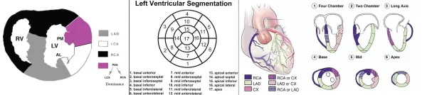
Coronary artery disease
Visualization of the coronary arteries and regional wall motion
CASE 1-1 Normal coronary arteries
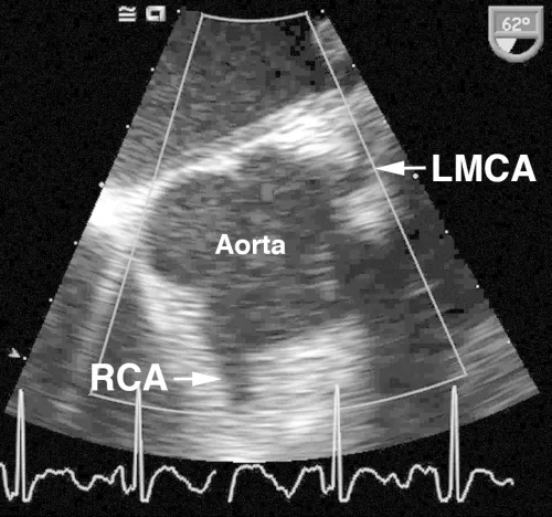
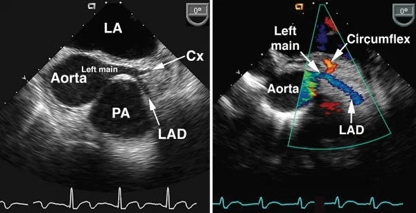
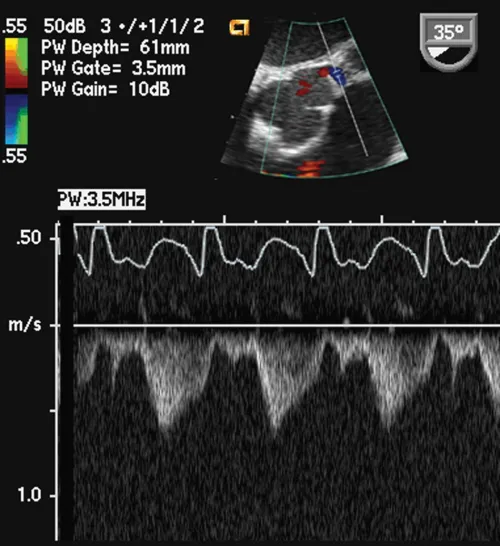
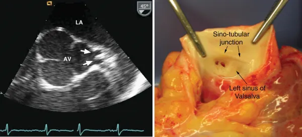
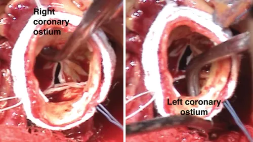
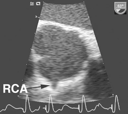
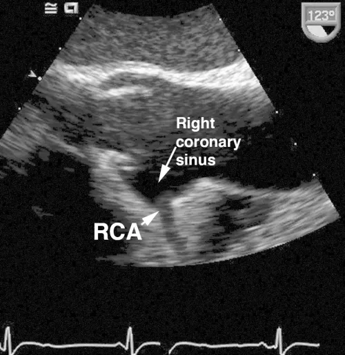
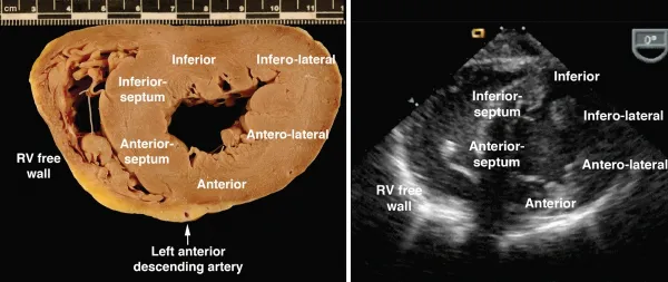
Comments
As shown in these examples, the proximal coronary arteries can often be visualized on TEE. The left main coronary artery arises from the left coronary sinus of Valsalva, is easily visualized in over 85% of patients and has a normal diameter of 4.2 ± 0.7 mm, with a slightly smaller average diameter in women (3.5 mm) compared with men (4.3 mm). The left main coronary artery bifurcates into the left anterior descendin...
Indice dei contenuti
Stili delle citazioni per Intraoperative and Interventional Echocardiography
APA 6 Citation
Oxorn, D., & Otto, C. (2016). Intraoperative and Interventional Echocardiography (2nd ed.). Elsevier Health Sciences. Retrieved from https://www.perlego.com/book/2938098/intraoperative-and-interventional-echocardiography-atlas-of-transesophageal-imaging-ebook-pdf (Original work published 2016)
Chicago Citation
Oxorn, Donald, and Catherine Otto. (2016) 2016. Intraoperative and Interventional Echocardiography. 2nd ed. Elsevier Health Sciences. https://www.perlego.com/book/2938098/intraoperative-and-interventional-echocardiography-atlas-of-transesophageal-imaging-ebook-pdf.
Harvard Citation
Oxorn, D. and Otto, C. (2016) Intraoperative and Interventional Echocardiography. 2nd edn. Elsevier Health Sciences. Available at: https://www.perlego.com/book/2938098/intraoperative-and-interventional-echocardiography-atlas-of-transesophageal-imaging-ebook-pdf (Accessed: 15 October 2022).
MLA 7 Citation
Oxorn, Donald, and Catherine Otto. Intraoperative and Interventional Echocardiography. 2nd ed. Elsevier Health Sciences, 2016. Web. 15 Oct. 2022.