eBook - ePub
Intraoperative and Interventional Echocardiography
Atlas of Transesophageal Imaging E-Book
Donald Oxorn, Catherine M. Otto
This is a test
- 448 pages
- English
- ePUB (adapté aux mobiles)
- Disponible sur iOS et Android
eBook - ePub
Intraoperative and Interventional Echocardiography
Atlas of Transesophageal Imaging E-Book
Donald Oxorn, Catherine M. Otto
Détails du livre
Aperçu du livre
Table des matières
Citations
À propos de ce livre
Case-based and heavily illustrated, Intraoperative and Interventional Echocardiography: Atlas of Transesophageal Imaging, 2nd Edition covers virtually every clinical scenario in which you're likely to use TEE. Drs. Donald C. Oxorn and Catherine M. Otto provide practical, how-to guidance on transesophageal echocardiography, including new approaches and state-of-the-art technologies. More than 1, 500 images sharpen your image acquisition and analysis skills and help you master this challenging technique.
- Real-world cases and abundant, detailed figures and tables show you exactly how to proceed, step by step, and get the best results.
- Each case begins with a brief presentation and discussion, and integrates clinical echocardiography, surgical pathology, and other imaging data.
- Clear descriptions of preoperative pathology guide you in choosing the best approach to common problems.
- The practice-based learning approach with expert commentary for each case helps you retain complex information and apply it in your daily practice.
- Every chapter has been thoroughly revised, with discussions of new technology and new techniques, including several techniques that are on the verge of becoming mainstream.
- New chapters cover current transcatheter valve therapies and device closures.
Foire aux questions
Comment puis-je résilier mon abonnement ?
Il vous suffit de vous rendre dans la section compte dans paramètres et de cliquer sur « Résilier l’abonnement ». C’est aussi simple que cela ! Une fois que vous aurez résilié votre abonnement, il restera actif pour le reste de la période pour laquelle vous avez payé. Découvrez-en plus ici.
Puis-je / comment puis-je télécharger des livres ?
Pour le moment, tous nos livres en format ePub adaptés aux mobiles peuvent être téléchargés via l’application. La plupart de nos PDF sont également disponibles en téléchargement et les autres seront téléchargeables très prochainement. Découvrez-en plus ici.
Quelle est la différence entre les formules tarifaires ?
Les deux abonnements vous donnent un accès complet à la bibliothèque et à toutes les fonctionnalités de Perlego. Les seules différences sont les tarifs ainsi que la période d’abonnement : avec l’abonnement annuel, vous économiserez environ 30 % par rapport à 12 mois d’abonnement mensuel.
Qu’est-ce que Perlego ?
Nous sommes un service d’abonnement à des ouvrages universitaires en ligne, où vous pouvez accéder à toute une bibliothèque pour un prix inférieur à celui d’un seul livre par mois. Avec plus d’un million de livres sur plus de 1 000 sujets, nous avons ce qu’il vous faut ! Découvrez-en plus ici.
Prenez-vous en charge la synthèse vocale ?
Recherchez le symbole Écouter sur votre prochain livre pour voir si vous pouvez l’écouter. L’outil Écouter lit le texte à haute voix pour vous, en surlignant le passage qui est en cours de lecture. Vous pouvez le mettre sur pause, l’accélérer ou le ralentir. Découvrez-en plus ici.
Est-ce que Intraoperative and Interventional Echocardiography est un PDF/ePUB en ligne ?
Oui, vous pouvez accéder à Intraoperative and Interventional Echocardiography par Donald Oxorn, Catherine M. Otto en format PDF et/ou ePUB ainsi qu’à d’autres livres populaires dans Medicina et Cardiología. Nous disposons de plus d’un million d’ouvrages à découvrir dans notre catalogue.
Informations
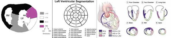
Chapter 1
Coronary artery disease
Visualization of the coronary arteries and regional wall motion
CASE 1-1 Normal coronary arteries
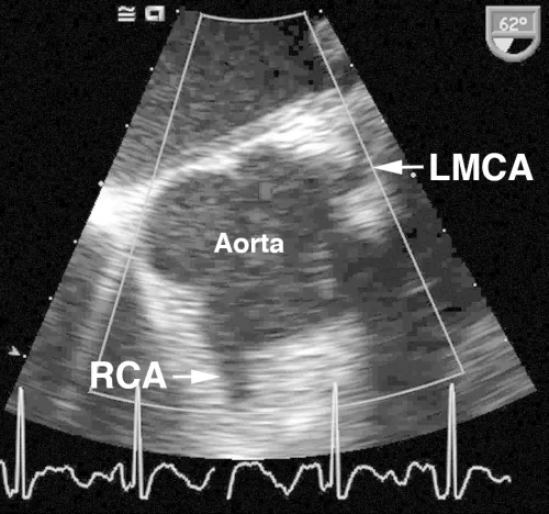
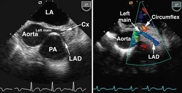
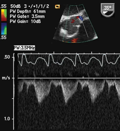
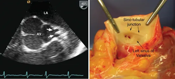
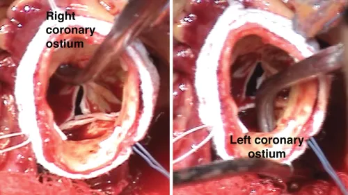
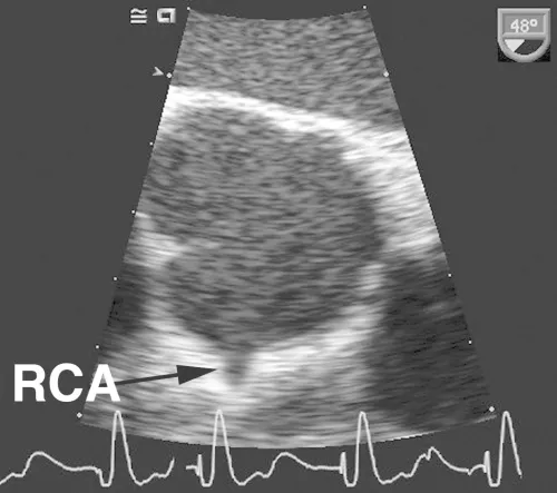
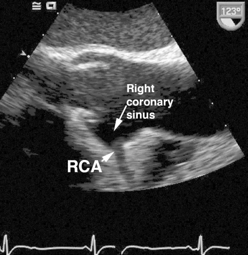
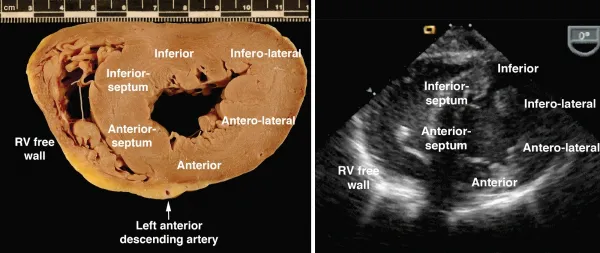
Comments
As shown in these examples, the proximal coronary arteries can often be visualized on TEE. The left main coronary artery arises from the left coronary sinus of Valsalva, is easily visualized in over 85% of patients and has a normal diameter of 4.2 ± 0.7 mm, with a slightly smaller average diameter in women (3.5 mm) compared with men (4.3 mm). The left main coronary artery bifurcates into the left anterior descendin...
Table des matières
Normes de citation pour Intraoperative and Interventional Echocardiography
APA 6 Citation
Oxorn, D., & Otto, C. (2016). Intraoperative and Interventional Echocardiography (2nd ed.). Elsevier Health Sciences. Retrieved from https://www.perlego.com/book/2938098/intraoperative-and-interventional-echocardiography-atlas-of-transesophageal-imaging-ebook-pdf (Original work published 2016)
Chicago Citation
Oxorn, Donald, and Catherine Otto. (2016) 2016. Intraoperative and Interventional Echocardiography. 2nd ed. Elsevier Health Sciences. https://www.perlego.com/book/2938098/intraoperative-and-interventional-echocardiography-atlas-of-transesophageal-imaging-ebook-pdf.
Harvard Citation
Oxorn, D. and Otto, C. (2016) Intraoperative and Interventional Echocardiography. 2nd edn. Elsevier Health Sciences. Available at: https://www.perlego.com/book/2938098/intraoperative-and-interventional-echocardiography-atlas-of-transesophageal-imaging-ebook-pdf (Accessed: 15 October 2022).
MLA 7 Citation
Oxorn, Donald, and Catherine Otto. Intraoperative and Interventional Echocardiography. 2nd ed. Elsevier Health Sciences, 2016. Web. 15 Oct. 2022.