eBook - ePub
Intraoperative and Interventional Echocardiography
Atlas of Transesophageal Imaging E-Book
Donald Oxorn, Catherine M. Otto
This is a test
- 448 Seiten
- English
- ePUB (handyfreundlich)
- Über iOS und Android verfügbar
eBook - ePub
Intraoperative and Interventional Echocardiography
Atlas of Transesophageal Imaging E-Book
Donald Oxorn, Catherine M. Otto
Angaben zum Buch
Buchvorschau
Inhaltsverzeichnis
Quellenangaben
Über dieses Buch
Case-based and heavily illustrated, Intraoperative and Interventional Echocardiography: Atlas of Transesophageal Imaging, 2nd Edition covers virtually every clinical scenario in which you're likely to use TEE. Drs. Donald C. Oxorn and Catherine M. Otto provide practical, how-to guidance on transesophageal echocardiography, including new approaches and state-of-the-art technologies. More than 1, 500 images sharpen your image acquisition and analysis skills and help you master this challenging technique.
- Real-world cases and abundant, detailed figures and tables show you exactly how to proceed, step by step, and get the best results.
- Each case begins with a brief presentation and discussion, and integrates clinical echocardiography, surgical pathology, and other imaging data.
- Clear descriptions of preoperative pathology guide you in choosing the best approach to common problems.
- The practice-based learning approach with expert commentary for each case helps you retain complex information and apply it in your daily practice.
- Every chapter has been thoroughly revised, with discussions of new technology and new techniques, including several techniques that are on the verge of becoming mainstream.
- New chapters cover current transcatheter valve therapies and device closures.
Häufig gestellte Fragen
Wie kann ich mein Abo kündigen?
Gehe einfach zum Kontobereich in den Einstellungen und klicke auf „Abo kündigen“ – ganz einfach. Nachdem du gekündigt hast, bleibt deine Mitgliedschaft für den verbleibenden Abozeitraum, den du bereits bezahlt hast, aktiv. Mehr Informationen hier.
(Wie) Kann ich Bücher herunterladen?
Derzeit stehen all unsere auf Mobilgeräte reagierenden ePub-Bücher zum Download über die App zur Verfügung. Die meisten unserer PDFs stehen ebenfalls zum Download bereit; wir arbeiten daran, auch die übrigen PDFs zum Download anzubieten, bei denen dies aktuell noch nicht möglich ist. Weitere Informationen hier.
Welcher Unterschied besteht bei den Preisen zwischen den Aboplänen?
Mit beiden Aboplänen erhältst du vollen Zugang zur Bibliothek und allen Funktionen von Perlego. Die einzigen Unterschiede bestehen im Preis und dem Abozeitraum: Mit dem Jahresabo sparst du auf 12 Monate gerechnet im Vergleich zum Monatsabo rund 30 %.
Was ist Perlego?
Wir sind ein Online-Abodienst für Lehrbücher, bei dem du für weniger als den Preis eines einzelnen Buches pro Monat Zugang zu einer ganzen Online-Bibliothek erhältst. Mit über 1 Million Büchern zu über 1.000 verschiedenen Themen haben wir bestimmt alles, was du brauchst! Weitere Informationen hier.
Unterstützt Perlego Text-zu-Sprache?
Achte auf das Symbol zum Vorlesen in deinem nächsten Buch, um zu sehen, ob du es dir auch anhören kannst. Bei diesem Tool wird dir Text laut vorgelesen, wobei der Text beim Vorlesen auch grafisch hervorgehoben wird. Du kannst das Vorlesen jederzeit anhalten, beschleunigen und verlangsamen. Weitere Informationen hier.
Ist Intraoperative and Interventional Echocardiography als Online-PDF/ePub verfügbar?
Ja, du hast Zugang zu Intraoperative and Interventional Echocardiography von Donald Oxorn, Catherine M. Otto im PDF- und/oder ePub-Format sowie zu anderen beliebten Büchern aus Medicina & Cardiología. Aus unserem Katalog stehen dir über 1 Million Bücher zur Verfügung.
Information
Chapter 1
Coronary artery disease
Visualization of the coronary arteries and regional wall motion
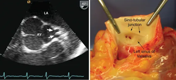
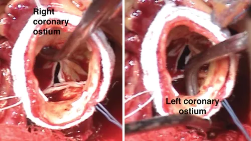
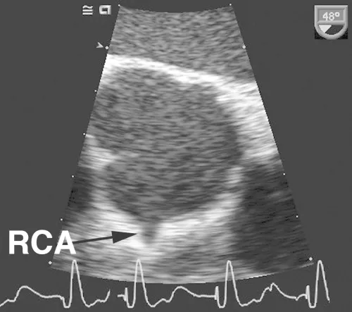
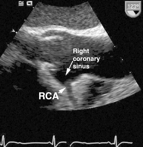
CASE 1-1 Normal coronary arteries
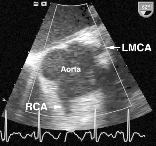
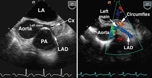
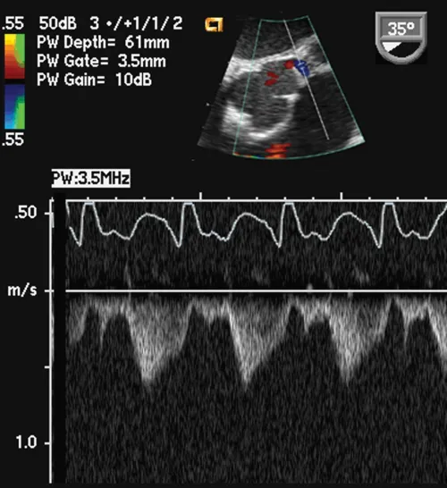
Comments
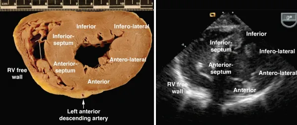
As shown in these examples, the proximal coronary arteries can often be visualized on TEE. The left main coronary artery arises from the left coronary sinus of Valsalva, is easily visualized in over 85% of patients and has a normal diameter of 4.2 ± 0.7 mm, with a slightly smaller average diameter in women (3.5 mm) compared with men (4.3 mm). The left main coronary artery bifurcates into the left anterior descendin...
Inhaltsverzeichnis
Zitierstile für Intraoperative and Interventional Echocardiography
APA 6 Citation
Oxorn, D., & Otto, C. (2016). Intraoperative and Interventional Echocardiography (2nd ed.). Elsevier Health Sciences. Retrieved from https://www.perlego.com/book/2938098/intraoperative-and-interventional-echocardiography-atlas-of-transesophageal-imaging-ebook-pdf (Original work published 2016)
Chicago Citation
Oxorn, Donald, and Catherine Otto. (2016) 2016. Intraoperative and Interventional Echocardiography. 2nd ed. Elsevier Health Sciences. https://www.perlego.com/book/2938098/intraoperative-and-interventional-echocardiography-atlas-of-transesophageal-imaging-ebook-pdf.
Harvard Citation
Oxorn, D. and Otto, C. (2016) Intraoperative and Interventional Echocardiography. 2nd edn. Elsevier Health Sciences. Available at: https://www.perlego.com/book/2938098/intraoperative-and-interventional-echocardiography-atlas-of-transesophageal-imaging-ebook-pdf (Accessed: 15 October 2022).
MLA 7 Citation
Oxorn, Donald, and Catherine Otto. Intraoperative and Interventional Echocardiography. 2nd ed. Elsevier Health Sciences, 2016. Web. 15 Oct. 2022.