Protein Bioinformatics
eBook - ePub

Protein Bioinformatics

From Sequence to Function

M. Michael Gromiha

  1. 339 Seiten
  2. English
  3. ePUB (handyfreundlich)
  4. Über iOS und Android verfügbar
eBook - ePub

Protein Bioinformatics

From Sequence to Function

M. Michael Gromiha

Angaben zum Buch
Buchvorschau
Inhaltsverzeichnis
Quellenangaben

Über dieses Buch

One of the most pressing tasks in biotechnology today is to unlock the function of each of the thousands of new genes identified every day. Scientists do this by analyzing and interpreting proteins, which are considered the task force of a gene. This single source reference covers all aspects of proteins, explaining fundamentals, synthesizing the latest literature, and demonstrating the most important bioinformatics tools available today for protein analysis, interpretation and prediction. Students and researchers of biotechnology, bioinformatics, proteomics, protein engineering, biophysics, computational biology, molecular modeling, and drug design will find this a ready reference for staying current and productive in this fast evolving interdisciplinary field.

  • Explains all aspects of proteins including sequence and structure analysis, prediction of protein structures, protein folding, protein stability, and protein interactions
  • Presents a cohesive and accessible overview of the field, using illustrations to explain key concepts and detailed exercises for students.

Häufig gestellte Fragen

Wie kann ich mein Abo kündigen?
Gehe einfach zum Kontobereich in den Einstellungen und klicke auf „Abo kündigen“ – ganz einfach. Nachdem du gekündigt hast, bleibt deine Mitgliedschaft für den verbleibenden Abozeitraum, den du bereits bezahlt hast, aktiv. Mehr Informationen hier.
(Wie) Kann ich Bücher herunterladen?
Derzeit stehen all unsere auf Mobilgeräte reagierenden ePub-Bücher zum Download über die App zur Verfügung. Die meisten unserer PDFs stehen ebenfalls zum Download bereit; wir arbeiten daran, auch die übrigen PDFs zum Download anzubieten, bei denen dies aktuell noch nicht möglich ist. Weitere Informationen hier.
Welcher Unterschied besteht bei den Preisen zwischen den Aboplänen?
Mit beiden Aboplänen erhältst du vollen Zugang zur Bibliothek und allen Funktionen von Perlego. Die einzigen Unterschiede bestehen im Preis und dem Abozeitraum: Mit dem Jahresabo sparst du auf 12 Monate gerechnet im Vergleich zum Monatsabo rund 30 %.
Was ist Perlego?
Wir sind ein Online-Abodienst für Lehrbücher, bei dem du für weniger als den Preis eines einzelnen Buches pro Monat Zugang zu einer ganzen Online-Bibliothek erhältst. Mit über 1 Million Büchern zu über 1.000 verschiedenen Themen haben wir bestimmt alles, was du brauchst! Weitere Informationen hier.
Unterstützt Perlego Text-zu-Sprache?
Achte auf das Symbol zum Vorlesen in deinem nächsten Buch, um zu sehen, ob du es dir auch anhören kannst. Bei diesem Tool wird dir Text laut vorgelesen, wobei der Text beim Vorlesen auch grafisch hervorgehoben wird. Du kannst das Vorlesen jederzeit anhalten, beschleunigen und verlangsamen. Weitere Informationen hier.
Ist Protein Bioinformatics als Online-PDF/ePub verfügbar?
Ja, du hast Zugang zu Protein Bioinformatics von M. Michael Gromiha im PDF- und/oder ePub-Format sowie zu anderen beliebten Büchern aus Scienze biologiche & Biologia molecolare. Aus unserem Katalog stehen dir über 1 Million Bücher zur Verfügung.

Information

Jahr
2011
ISBN
9780123884244
Chapter 1

Proteins

Publisher Summary

This chapter describes the functional properties of proteins. The functional properties of proteins depend on their three-dimensional structures. The native structure of a protein can be experimentally determined using X-ray crystallography, nuclear magnetic resonance (NMR) spectroscopy, electron microscopy, etc. Proteins perform a variety of functions, including enzymatic catalysis, transporting ions and molecules from one organ to another, nutrients, contractile system of muscles, tendons, cartilage, antibodies, and regulating cellular and physiological activities. Deciphering the three-dimensional structure of a protein from its amino acid sequence is a long-standing goal in molecular and computational biology. A protein chain is formed by several amino acids in which the amino group of the first amino acid and the carboxyl group of the last amino acid remain intact, and the chain is said to extend from the amino (N) to the carboxyl (C) terminus. This chain of amino acids is called a polypeptide chain, main chain, or backbone. These polypeptide chains that have specific functions are called proteins. In a polypeptide chain, the -carbon atoms of adjacent amino acids are separated by three covalent bonds arranged as Ca—C—N—Ca. Proteins are broadly classified into two major groups: fibrous proteins, having polypeptide chains arranged in long strands, and globular proteins, with polypeptide chains folded into a spherical or globular shape.
Proteins perform a variety of functions, including enzymatic catalysis, transporting ions and molecules from one organ to another, nutrients, contractile system of muscles, tendons, cartilage, antibodies, and regulating cellular and physiological activities. The functional properties of proteins depend on their three-dimensional structures. The native structure of a protein can be experimentally determined using X-ray crystallography, nuclear magnetic resonance (NMR) spectroscopy, electron microscopy, etc. Over the past 40 years, the structures of more than 53,000 proteins (as of May 12, 2009) have been determined. On the other hand, the amino acid sequences are determined for more than eight million proteins (as of May 5, 2009). The specific sequence of amino acids in a polypeptide chain folds to generate compact domains with a particular three-dimensional structure. Anfinsen (1973) stated that the polypeptide chain itself contains all the information necessary to specify its three-dimensional structure. Deciphering the three-dimensional structure of a protein from its amino acid sequence is a long-standing goal in molecular and computational biology.

1.1 Building blocks

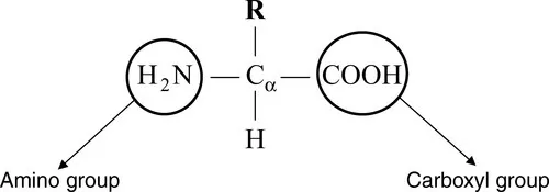
Protein sequences consist of 20 different kinds of chemical compounds, known as amino acids, and they serve as building blocks of proteins. Amino acids contain a central carbon atom (Cα), which is attached to a hydrogen atom, an amino group (NH2), and a carboxyl group (COOH) as shown in Figure 1.1. The letter R in Figure 1.1 indicates the presence of a side chain, which distinguishes each amino acid.
f01-01-9788131222973
Figure 1.1 Representation of amino acids. R is the side chain that varies for the 20 amino acids.

1.1.1 Amino acids

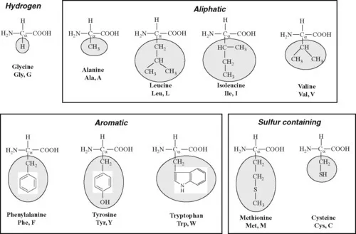
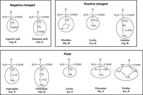
Amino acids are naturally of 20 different types as specified by the genetic code emerged from DNA sequences. Furthermore, nonnatural amino acids occur, in rare cases,as the products of enzymatic modifications after translocation. The major difference among the 20 amino acids is the side chain attached to the Cα through its fourth valance. The variation of side chains in 20 amino acids is shown in Figure 1.2. These residues are represented by conventional three- and one-letter codes. Most of the databases use single-letter codes.
f01-02a-9788131222973
f01-02b-9788131222973
Figure 1.2 The common 20 amino acids in proteins. The three- and one-letter codes for the amino acids are also given. The amino acids are classified into hydrophobic (hydrogen, aliphatic, aromatic, and sulfur containing) and hydrophilic (negatively charged, positively charged, and polar). The side chains are marked with oval boxes.
The amino acids are broadly divided into two groups, hydrophobic and hydrophilic, based on the tendency of their interactions in the presence of water molecule. The hydrophobic residues have the tendency of adhering to one another in aqueous environment. Generally, amino acids, Ala (A), Cys (C), Phe (F), Gly (G), Ile (I), Leu (L), Met (M), Val (V), Trp (W), and Tyr (Y), are considered as hydrophobic residues. In this category, Ala, Ile, Leu, and Val contain aliphatic side chains; Phe, Trp,and Tyr contain aromatic side chains; and Cys and Met contain sulfur atom. Gly has no side chain, and it has hydrogen (H) at the fourth position. Two Cys residues in different parts of the polypeptide chain but adjacent to each other in the threedimensional structure of a protein can be oxidized to form a disulfide bridge. The formation of disulfide bridges in protein structures stabilizes the protein, making it less susceptible to degradation.
Amino acids, Asp (D), Glu (E), His (H), Lys (K), Asn (N), Pro (P), Gln (Q), Arg (R), Ser (S), and Thr (T), are classified as hydrophilic residues. In this category, Asp and Glu are negatively charged; His, Lys, and Arg are positively charged; and others are polar and uncharged.

1.1.2 Formation of peptide bonds

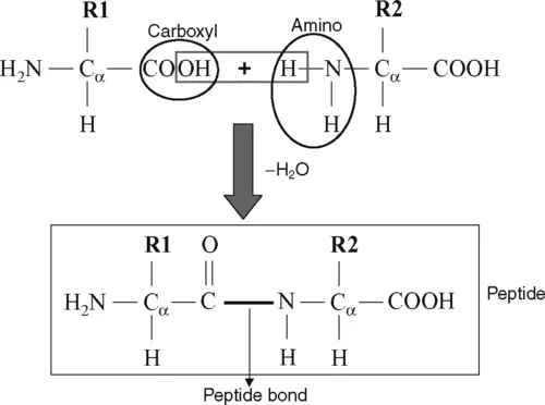
The carboxyl group of one amino acid interacts with the amino group of another to form a peptide bond by the elimination of water (Figure 1.3). Amino acids are joined end-to-end during protein synthesis by the formation of such peptide bonds. The peptide bond (C
glyph_sbnd
N) has a partial double-bond character due to resonance, and hence there is no rotation about the peptide bond. In Figure 1.3, the peptide is represented as a planar unit with the C
glyph_dbnd
O and N
glyph_sbnd
H groups positioning in opposite directions in the plane. This is called trans-peptide. There is another form, cis-peptide in which the C
glyph_dbnd
O and N
glyph_sbnd
H groups point in the same direction. To avoid steric hindrance, the trans form is frequently presented in protein structures for all amino acids except Pro, which has both trans and cis forms. The cis prolines are found in bends of the polypeptide chains.
f01-03-9788131222973
Figure 1.3 Formation of a peptide bond by the elimination of a water molecule.
A protein chain is formed by several amino acids in which the amino group of the first amino acid and the carboxyl group of the last amino acid remain intact, and the chain is said to extend from the amino (N) to the carboxyl (C) terminus. This chain of amino acids is called a polypeptide chain, main chain, or backbone. Amino acids in a polypeptide chain lack a hydrogen atom at the amino terminal and an OH group at the carboxyl terminal (except at the ends), and hence amino acids are also called amino acid residues (simply residues). Nature selects the combination of amino acid residues to form polypeptide chains for their function, similar to the combination of alpha...

Inhaltsverzeichnis

Zitierstile für Protein Bioinformatics

APA 6 Citation

Gromiha, M. (2011). Protein Bioinformatics ([edition unavailable]). Elsevier Science. Retrieved from https://www.perlego.com/book/1836074/protein-bioinformatics-from-sequence-to-function-pdf (Original work published 2011)

Chicago Citation

Gromiha, Michael. (2011) 2011. Protein Bioinformatics. [Edition unavailable]. Elsevier Science. https://www.perlego.com/book/1836074/protein-bioinformatics-from-sequence-to-function-pdf.

Harvard Citation

Gromiha, M. (2011) Protein Bioinformatics. [edition unavailable]. Elsevier Science. Available at: https://www.perlego.com/book/1836074/protein-bioinformatics-from-sequence-to-function-pdf (Accessed: 15 October 2022).

MLA 7 Citation

Gromiha, Michael. Protein Bioinformatics. [edition unavailable]. Elsevier Science, 2011. Web. 15 Oct. 2022.