Sturkie's Avian Physiology
eBook - ePub

Sturkie's Avian Physiology

G. Causey Whittow, G. Causey Whittow

Buch teilen
  1. 704 Seiten
  2. English
  3. ePUB (handyfreundlich)
  4. Über iOS und Android verfügbar
eBook - ePub

Sturkie's Avian Physiology

G. Causey Whittow, G. Causey Whittow

Angaben zum Buch
Buchvorschau
Inhaltsverzeichnis
Quellenangaben

Über dieses Buch

Sturkie's Avian Physiology is the classic comprehensive single volume on the physiology of domestic as well as wild birds. The Fifth Edition is thoroughly revised and updated, and includes new chapters on the physiology of incubation and growth. Chapters on the nervous system and sensory organs have been greatly expanded due to the many recent advances in the field. The text also covers the physiology of flight, reproduction in both male and female birds, and the immunophysiology of birds.

The Fifth Edition, like the earlier editions, is a must for anyone interested in comparative physiology, poultry science, veterinary medicine, and related fields. This volume establishes the standard for those who need the latest and best information on the physiology of birds.

  • Thoroughly updated and revised
  • Coverage of both domestic and wild birds
  • New larger format
  • Only comprehensive, single volume devoted to birds

Häufig gestellte Fragen

Wie kann ich mein Abo kündigen?
Gehe einfach zum Kontobereich in den Einstellungen und klicke auf „Abo kündigen“ – ganz einfach. Nachdem du gekündigt hast, bleibt deine Mitgliedschaft für den verbleibenden Abozeitraum, den du bereits bezahlt hast, aktiv. Mehr Informationen hier.
(Wie) Kann ich Bücher herunterladen?
Derzeit stehen all unsere auf Mobilgeräte reagierenden ePub-Bücher zum Download über die App zur Verfügung. Die meisten unserer PDFs stehen ebenfalls zum Download bereit; wir arbeiten daran, auch die übrigen PDFs zum Download anzubieten, bei denen dies aktuell noch nicht möglich ist. Weitere Informationen hier.
Welcher Unterschied besteht bei den Preisen zwischen den Aboplänen?
Mit beiden Aboplänen erhältst du vollen Zugang zur Bibliothek und allen Funktionen von Perlego. Die einzigen Unterschiede bestehen im Preis und dem Abozeitraum: Mit dem Jahresabo sparst du auf 12 Monate gerechnet im Vergleich zum Monatsabo rund 30 %.
Was ist Perlego?
Wir sind ein Online-Abodienst für Lehrbücher, bei dem du für weniger als den Preis eines einzelnen Buches pro Monat Zugang zu einer ganzen Online-Bibliothek erhältst. Mit über 1 Million Büchern zu über 1.000 verschiedenen Themen haben wir bestimmt alles, was du brauchst! Weitere Informationen hier.
Unterstützt Perlego Text-zu-Sprache?
Achte auf das Symbol zum Vorlesen in deinem nächsten Buch, um zu sehen, ob du es dir auch anhören kannst. Bei diesem Tool wird dir Text laut vorgelesen, wobei der Text beim Vorlesen auch grafisch hervorgehoben wird. Du kannst das Vorlesen jederzeit anhalten, beschleunigen und verlangsamen. Weitere Informationen hier.
Ist Sturkie's Avian Physiology als Online-PDF/ePub verfügbar?
Ja, du hast Zugang zu Sturkie's Avian Physiology von G. Causey Whittow, G. Causey Whittow im PDF- und/oder ePub-Format sowie zu anderen beliebten Büchern aus Biological Sciences & Zoology. Aus unserem Katalog stehen dir über 1 Million Bücher zur Verfügung.

Information

Jahr
1999
ISBN
9780080542089
CHAPTER 1 Sensory Physiology: Vision
ONUR. GÜNTÜRKÜN
AE Biopsychologie, Fakultät für Psychologie, Ruhr-Universität Bochum, 44780 Bochum, Germany
I. Introduction
II. Structure and Functions of the Eye
A. Eye Shape, Stereopsis, and Acuity
B. Retina
III. Central Processing—Anatomy and Function
A. Centrifugal Pathway
B. Tectofugal Pathway
C. Thalamofugal Pathway 12 References

I INTRODUCTION

Birds are the most visually dependent class of vertebrates and the phrase of Rochon-Duvigneaud (1943) that a pigeon is nothing else but two eyes with wings is probably valid for most avian species. Man, a highly visual primate, sees the world with the information transmitted by about one million fibers within each of his optic nerves. This is only 40% of the number of retinal axons counted in a single optic nerve of pigeons and chicks (Binggeli and Paule, 1969; Rager and Rager, 1978). The acuity of many birds of prey surpasses that of other living beings (Fox et al., 1976) and even the unspecialized pigeon excels relative to humans in its ability to discriminate luminances (Hodos et al., 1985) and discern subtle color differences (Emmerton and Delius, 1980). Food-storing birds like Clark’s nutcracker store 33,000 seeds in about 6,600 caches to survive in winter (Vander Wall and Balda, 1977). Pigeons acquire visual concepts of, for example, “animals” (Roberts and Mazmanian, 1988), “same versus different” (Wright et al., 1988), and even cartoon figures such as “Charlie Brown” (Cerella, 1980). They communicate using visual symbols (Lubinski and MacCorquodale, 1984) and are able to rank optic patterns by using transitive inference logic (von Fersen et al., 1992). If we, on the basis of countless evidence, assume that the visual system of amniotes has evolved only once (Shimizu and Karten, 1993), the avian visual system is a remarkable model to explore its morphology, its modes of operations, and the unanticipated complexity of its function.

II STRUCTURE AND FUNCTIONS OF THE EYE

Avian eyes take up a considerable volume of the bird’s head and are very large in relation to brain size (Figure 1). In general terms, the structure of their eyes is not much different from that of other vertebrates. Incoming light has to pass through four media: the cornea, the anterior chamber, the lens, and the vitrous body, before reaching the retina, where photoreceptors convert light energy into electric impulses by bleaching of visual pigments. All four optic media are remarkably transparent, transmitting wavelengths down to at least 310 nm in the near-ultraviolet range (Emmerton et al., 1980).
image
FIGURE 1 Drawing of a horizontal section of the chicken eye showing the position of the eyes within the head.
(From H. Evans, 1996.)
The avian retina is completely avascularized to prevent shadows and light scattering. This arrangement is associated with the presence of an unusual nutritional device specific for birds—the pecten. This black pigmented and manifoldly pleated structure projects from the ventral retina above the exit of the optic nerve toward the lens and is completely made up of blood vessels and extravascular pigmented stromal cells. There is evidence that it also has a nutritive function. This is shown by the presence of an oxygen gradient from the pecten to the retina, the passing of nutrients from the pecten into the vitreous, and the observation that fluorescent markers pass from the pecten into the vitreous (Bellhorn and Bellhorn, 1975). Also, Pettigrew et al. (1990) posit that the inertia of the pecten during saccadic eye movements could be used like a shaker to propel oxygen and nutrients within the eye.

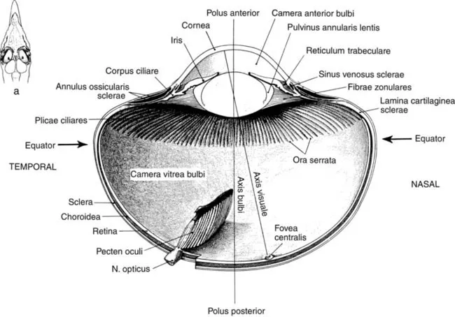
A Eye Shape, Stereopsis, and Acuity

The eyeshapes of birds are a result of ecological requirements (Figure 2). Generally, acuity can be maximized by increasing the anterior focal length of an eye; the optic image is then spread over a larger retinal surface and thus over a larger number of photoreceptors (Martin, 1993). Increasing the number of photoreceptors also makes it possible to connect several receptors to single bipolar cells and thus to maximize visual detection even under low light conditions. Since an increase in eye size is advantageous, birds, which rely heavily on vision, generally have the largest absolute and relative eyes within the animal kingdom. The eye of the ostrich, for example, has an axial length of 50 mm, the largest of any land vertebrate and twice that of the human eye (Walls, 1942). The tube-shaped eyecups of birds of prey, which create an extremely large image on the retina, represent another extreme version of biological optimization to achieve high acuity. These eyes generally also have a low retinal convergence ratio (receptors per ganglion cell) so that the receptor inputs are not pooled to increase visual resolution (Snyder et al., 1977). However, these optimizations are limited by trade-offs for brightness sensitivity. Retinae in which receptors are not pooled function only optimally at high light intensities and, indeed, resolution of birds of prey deteriorates at dusk (Reymond, 1985).
image
FIGURE 2 Horizontal section through the head of the black-capped chickedee (Parus atricapillus) and the great owl (Bubo virginianus).
(From Perception and Motor Control in Birds, Form and function in the optical structure of bird eyes, G. R. Martin, pp. 5–34, Fig. 1.2, 1994, © Springer-Verlag.)
Visual acuity measurements in pigeons (Columba livia) have shown that the acuity in the frontal field depends on stimulus time (Bloch and Martinoya, 1982), wavelength of light (Hodos and Leibowitz, 1977), luminance (Hodos et al., 1976; Hodos and Leibowitz, 1977), and age of the pigeon (Hodos et al., 1991a). Under favorable conditions 1-year-old pigeons reach a frontal acuity of 12.7 c/deg, increase this value to 16–18 c/deg at 2 years, and decline to 3 c/deg at 17 years (Hodos et al., 1985, 1991b). The frontal binocular visual field of pigeons is represented in the superiotemporal area dorsalis, while the lateral monocular visual field is observed via the area centralis (both lack a true foveal depression). These two retinal regions seem to subserve different visual functions with differing capacities for optic resolutions. Behavioral studies show that many avian species, including pigeons, fixate distant objects preferentially with their lateral and monocular field (pigeon: Blough, 1971; dove: Friedman, 1975; kestrel: Fox et al., 1976; eagle: Reymond, 1985; passerine birds: Bischof, 1988; Kirmse, 1990). This behavior is often pronounced; birds orient themselves sideways in order to achieve a lateral orientation to the inspected object. This behavior, together with the fact that retinal ganglion cell densities reach peak values in the central fovea, suggest that resolution is maximal in the lateral visual field. However, the acuity of young pigeons is 12.6 c/deg in their lateral visual field and thus identical with the values obtained for frontal vision in same aged subjects (Hahmann and Güntürkü...

Inhaltsverzeichnis