
eBook - ePub
Pathology of Melanocytic Tumors E-Book
Klaus J. Busam, Richard A Scolyer, Pedram Gerami
This is a test
Buch teilen
- 608 Seiten
- English
- ePUB (handyfreundlich)
- Über iOS und Android verfügbar
eBook - ePub
Pathology of Melanocytic Tumors E-Book
Klaus J. Busam, Richard A Scolyer, Pedram Gerami
Angaben zum Buch
Buchvorschau
Inhaltsverzeichnis
Quellenangaben
Über dieses Buch
Constituting a large percentage of everyday diagnostic practice, melanocytic pathology is a complex and challenging area with many difficult-to-diagnose lesions. This highly illustrated reference, written by three of the world's leading dermatopathologists, provides authoritative guidance in the accurate diagnosis of even the most challenging pigmented skin tumors, helping you avoid pitfalls and recognize mimics.
- Covers nearly every variant of melanocytic tumors you're likely to see.
- Emphasizes how to arrive at an efficient, accurate diagnosis, and includes dermoscopic findings for optimal diagnostic precision.
- Discusses modern analytic techniques ( cytogenetics, molecular studies ) and how to use them for diagnosis.
- Includes numerous case examples to illustrate the differential diagnoses and work-up; how to use ancillary techniques, along with their pros, cons, and limitations; and clinical follow-up.
- Presents the knowledge and experience of Klaus Busam, Pedram Gerami, and Richard Scolyer, – three dermatopathologists who are globally renowned for their expertise in melanoma pathology and analysis of melanocytic tumors by modern ancillary diagnostic techniques.
Häufig gestellte Fragen
Wie kann ich mein Abo kündigen?
Gehe einfach zum Kontobereich in den Einstellungen und klicke auf „Abo kündigen“ – ganz einfach. Nachdem du gekündigt hast, bleibt deine Mitgliedschaft für den verbleibenden Abozeitraum, den du bereits bezahlt hast, aktiv. Mehr Informationen hier.
(Wie) Kann ich Bücher herunterladen?
Derzeit stehen all unsere auf Mobilgeräte reagierenden ePub-Bücher zum Download über die App zur Verfügung. Die meisten unserer PDFs stehen ebenfalls zum Download bereit; wir arbeiten daran, auch die übrigen PDFs zum Download anzubieten, bei denen dies aktuell noch nicht möglich ist. Weitere Informationen hier.
Welcher Unterschied besteht bei den Preisen zwischen den Aboplänen?
Mit beiden Aboplänen erhältst du vollen Zugang zur Bibliothek und allen Funktionen von Perlego. Die einzigen Unterschiede bestehen im Preis und dem Abozeitraum: Mit dem Jahresabo sparst du auf 12 Monate gerechnet im Vergleich zum Monatsabo rund 30 %.
Was ist Perlego?
Wir sind ein Online-Abodienst für Lehrbücher, bei dem du für weniger als den Preis eines einzelnen Buches pro Monat Zugang zu einer ganzen Online-Bibliothek erhältst. Mit über 1 Million Büchern zu über 1.000 verschiedenen Themen haben wir bestimmt alles, was du brauchst! Weitere Informationen hier.
Unterstützt Perlego Text-zu-Sprache?
Achte auf das Symbol zum Vorlesen in deinem nächsten Buch, um zu sehen, ob du es dir auch anhören kannst. Bei diesem Tool wird dir Text laut vorgelesen, wobei der Text beim Vorlesen auch grafisch hervorgehoben wird. Du kannst das Vorlesen jederzeit anhalten, beschleunigen und verlangsamen. Weitere Informationen hier.
Ist Pathology of Melanocytic Tumors E-Book als Online-PDF/ePub verfügbar?
Ja, du hast Zugang zu Pathology of Melanocytic Tumors E-Book von Klaus J. Busam, Richard A Scolyer, Pedram Gerami im PDF- und/oder ePub-Format sowie zu anderen beliebten Büchern aus Medizin & Pathologie. Aus unserem Katalog stehen dir über 1 Million Bücher zur Verfügung.
Information
Thema
MedizinThema
PathologieSection I
Benign Cutaneous Melanocytic Proliferations
Introduction
- 1. Melanotic Macules, 2
- Klaus J. Busam
- 2. Acquired Melanocytic Nevi, 8
- Pedram Gerami and Klaus J. Busam
- 3. Congenital Melanocytic Nevi, 26
- Pedram Gerami
- 4. Spitz Nevi, 37
- Pedram Gerami and Klaus J. Busam
- 5. Blue Nevi and Dermal Melanocytosis, 61
- Klaus J. Busam
- 6. Deep Penetrating Nevi, 80
- Klaus J. Busam, Iwei Yeh, and Richard A. Scolyer
- 7. Melanocytic Nevi of Special Sites, 90
- Timothy VandenBoom and Pedram Gerami
- 8. Persistant/Recurrent Melanocytic Nevi, Traumatized Nevi, and Nevi Changing Under Therapy, 101
- Maija Kiuru, Pedram Gerami, and Klaus J. Busam
- 9. Combined Melanocytic Nevi, 112
- Klaus J. Busam and Richard A. Scolyer
- 10. Pigmented Epithelioid Melanocytoma, 124
- Artur Zembowicz, Jarish N. Cohen, and Philip E. LeBoit
1
Melanotic Macules
Klaus J. Busam
The term melanotic macule is used to refer to benign flat pigmented lesions, which are histopathologically characterized by melanin pigment deposited in basilar keratinocytes without or at times with a slight increase in the density of solitary units of junctional melanocytes. Cutaneous and mucosal melanotic macules are often labeled lentigo. The conjunctival equivalent has historically been termed acquired melanosis. In contrast to melanocytic nevi, there are no junctional nests in melanotic macules, lentigines, or benign acquired melanosis. The lesions differ from melanoma in situ clinically by a tendency toward small size and sharp circumscription and histopathologically by a low density of melanocytes. Melanotic macules are a common clinical presentation. They are benign and do not need to be treated. However, they may be biopsied when they display irregular features, are new, or are changing, to exclude melanoma in situ.
Solar Lentigo
Clinical Features
Solar lentigo manifests as a discrete pigmented macule or patch on sun-exposed skin of middle-aged or elderly individuals (Box 1.1).1 Its color may range from light tan to dark brown. A lesion's color may be uniform or heterogeneous. The size of a solar lentigo may be small (2 mm) or large (>1 cm). Its peripheral borders may be regular or ill-defined (Figs. 1.1 and 1.2). Multiple lesions of solar lentigo may coexist in the same anatomic region. They may collide with each other or with seborrheic or actinic keratoses, become confluent, and appear clinically complex. Clinical atypical lesions, which stand out from the background, prompt concerns for lentigo maligna (melanoma in situ) and are usually biopsied to assess for or exclude melanoma. Solar lentigines become more frequent and/or larger in area with age. Lesions of solar lentigo can also be found in young patients with sun-damaged skin and are common in children with xeroderma pigmentosum.
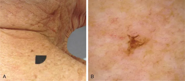
Fig. 1.1 Clinical Appearance of Solar Lentigo. (A) Pigmented macule. (B) This lesion is small but irregular in outline. Its asymmetric silhouette was the reason why it was biopsied.
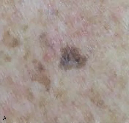
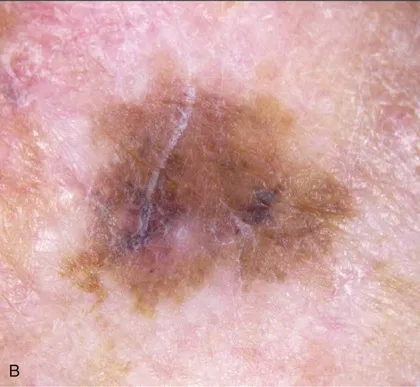
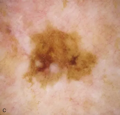
Fig. 1.2 Clinical Appearance of Solar Lentigo. (A) Hyperpigmented macule in a background of light brown macules on the chest of an elderly woman. (B) The lesion displays some irregular borders and variation in pigment intensity. (C) Corresponding dermoscopic findings.
A variant of solar lentigo clinically characterized by presentation as small dark macules, especially on the upper back, has been referred to as “ink spot” lentigo or reticulated melanotic macule.2
Histopathologic Findings
Solar lentigo is histopathologically characterized by hypermelanization of basilar keratinocytes (Figs. 1.3–1.5). Often there is associated slight epidermal hyperplasia resulting in elongated, club-shaped rete ridges. As rete ridges become longer, they may form a reticulate pattern. Lesions of solar lentigo may be associated with or develop into a seborrheic keratosis (Fig. 1.6). Pigmentation of keratinocytes is then typically most pronounced at the tip of rete ridges. In some lesions the rete ridges may...