
eBook - ePub
Pathology of Melanocytic Tumors E-Book
Klaus J. Busam, Richard A Scolyer, Pedram Gerami
This is a test
Partager le livre
- 608 pages
- English
- ePUB (adapté aux mobiles)
- Disponible sur iOS et Android
eBook - ePub
Pathology of Melanocytic Tumors E-Book
Klaus J. Busam, Richard A Scolyer, Pedram Gerami
Détails du livre
Aperçu du livre
Table des matières
Citations
À propos de ce livre
Constituting a large percentage of everyday diagnostic practice, melanocytic pathology is a complex and challenging area with many difficult-to-diagnose lesions. This highly illustrated reference, written by three of the world's leading dermatopathologists, provides authoritative guidance in the accurate diagnosis of even the most challenging pigmented skin tumors, helping you avoid pitfalls and recognize mimics.
- Covers nearly every variant of melanocytic tumors you're likely to see.
- Emphasizes how to arrive at an efficient, accurate diagnosis, and includes dermoscopic findings for optimal diagnostic precision.
- Discusses modern analytic techniques ( cytogenetics, molecular studies ) and how to use them for diagnosis.
- Includes numerous case examples to illustrate the differential diagnoses and work-up; how to use ancillary techniques, along with their pros, cons, and limitations; and clinical follow-up.
- Presents the knowledge and experience of Klaus Busam, Pedram Gerami, and Richard Scolyer, – three dermatopathologists who are globally renowned for their expertise in melanoma pathology and analysis of melanocytic tumors by modern ancillary diagnostic techniques.
Foire aux questions
Comment puis-je résilier mon abonnement ?
Il vous suffit de vous rendre dans la section compte dans paramètres et de cliquer sur « Résilier l’abonnement ». C’est aussi simple que cela ! Une fois que vous aurez résilié votre abonnement, il restera actif pour le reste de la période pour laquelle vous avez payé. Découvrez-en plus ici.
Puis-je / comment puis-je télécharger des livres ?
Pour le moment, tous nos livres en format ePub adaptés aux mobiles peuvent être téléchargés via l’application. La plupart de nos PDF sont également disponibles en téléchargement et les autres seront téléchargeables très prochainement. Découvrez-en plus ici.
Quelle est la différence entre les formules tarifaires ?
Les deux abonnements vous donnent un accès complet à la bibliothèque et à toutes les fonctionnalités de Perlego. Les seules différences sont les tarifs ainsi que la période d’abonnement : avec l’abonnement annuel, vous économiserez environ 30 % par rapport à 12 mois d’abonnement mensuel.
Qu’est-ce que Perlego ?
Nous sommes un service d’abonnement à des ouvrages universitaires en ligne, où vous pouvez accéder à toute une bibliothèque pour un prix inférieur à celui d’un seul livre par mois. Avec plus d’un million de livres sur plus de 1 000 sujets, nous avons ce qu’il vous faut ! Découvrez-en plus ici.
Prenez-vous en charge la synthèse vocale ?
Recherchez le symbole Écouter sur votre prochain livre pour voir si vous pouvez l’écouter. L’outil Écouter lit le texte à haute voix pour vous, en surlignant le passage qui est en cours de lecture. Vous pouvez le mettre sur pause, l’accélérer ou le ralentir. Découvrez-en plus ici.
Est-ce que Pathology of Melanocytic Tumors E-Book est un PDF/ePUB en ligne ?
Oui, vous pouvez accéder à Pathology of Melanocytic Tumors E-Book par Klaus J. Busam, Richard A Scolyer, Pedram Gerami en format PDF et/ou ePUB ainsi qu’à d’autres livres populaires dans Medizin et Pathologie. Nous disposons de plus d’un million d’ouvrages à découvrir dans notre catalogue.
Informations
Sujet
MedizinSous-sujet
PathologieSection I
Benign Cutaneous Melanocytic Proliferations
Introduction
- 1. Melanotic Macules, 2
- Klaus J. Busam
- 2. Acquired Melanocytic Nevi, 8
- Pedram Gerami and Klaus J. Busam
- 3. Congenital Melanocytic Nevi, 26
- Pedram Gerami
- 4. Spitz Nevi, 37
- Pedram Gerami and Klaus J. Busam
- 5. Blue Nevi and Dermal Melanocytosis, 61
- Klaus J. Busam
- 6. Deep Penetrating Nevi, 80
- Klaus J. Busam, Iwei Yeh, and Richard A. Scolyer
- 7. Melanocytic Nevi of Special Sites, 90
- Timothy VandenBoom and Pedram Gerami
- 8. Persistant/Recurrent Melanocytic Nevi, Traumatized Nevi, and Nevi Changing Under Therapy, 101
- Maija Kiuru, Pedram Gerami, and Klaus J. Busam
- 9. Combined Melanocytic Nevi, 112
- Klaus J. Busam and Richard A. Scolyer
- 10. Pigmented Epithelioid Melanocytoma, 124
- Artur Zembowicz, Jarish N. Cohen, and Philip E. LeBoit
1
Melanotic Macules
Klaus J. Busam
The term melanotic macule is used to refer to benign flat pigmented lesions, which are histopathologically characterized by melanin pigment deposited in basilar keratinocytes without or at times with a slight increase in the density of solitary units of junctional melanocytes. Cutaneous and mucosal melanotic macules are often labeled lentigo. The conjunctival equivalent has historically been termed acquired melanosis. In contrast to melanocytic nevi, there are no junctional nests in melanotic macules, lentigines, or benign acquired melanosis. The lesions differ from melanoma in situ clinically by a tendency toward small size and sharp circumscription and histopathologically by a low density of melanocytes. Melanotic macules are a common clinical presentation. They are benign and do not need to be treated. However, they may be biopsied when they display irregular features, are new, or are changing, to exclude melanoma in situ.
Solar Lentigo
Clinical Features
Solar lentigo manifests as a discrete pigmented macule or patch on sun-exposed skin of middle-aged or elderly individuals (Box 1.1).1 Its color may range from light tan to dark brown. A lesion's color may be uniform or heterogeneous. The size of a solar lentigo may be small (2 mm) or large (>1 cm). Its peripheral borders may be regular or ill-defined (Figs. 1.1 and 1.2). Multiple lesions of solar lentigo may coexist in the same anatomic region. They may collide with each other or with seborrheic or actinic keratoses, become confluent, and appear clinically complex. Clinical atypical lesions, which stand out from the background, prompt concerns for lentigo maligna (melanoma in situ) and are usually biopsied to assess for or exclude melanoma. Solar lentigines become more frequent and/or larger in area with age. Lesions of solar lentigo can also be found in young patients with sun-damaged skin and are common in children with xeroderma pigmentosum.
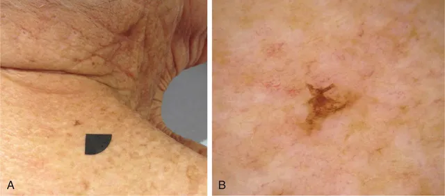
Fig. 1.1 Clinical Appearance of Solar Lentigo. (A) Pigmented macule. (B) This lesion is small but irregular in outline. Its asymmetric silhouette was the reason why it was biopsied.
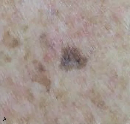
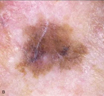
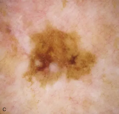
Fig. 1.2 Clinical Appearance of Solar Lentigo. (A) Hyperpigmented macule in a background of light brown macules on the chest of an elderly woman. (B) The lesion displays some irregular borders and variation in pigment intensity. (C) Corresponding dermoscopic findings.
A variant of solar lentigo clinically characterized by presentation as small dark macules, especially on the upper back, has been referred to as “ink spot” lentigo or reticulated melanotic macule.2
Histopathologic Findings
Solar lentigo is histopathologically characterized by hypermelanization of basilar keratinocytes (Figs. 1.3–1.5). Often there is associated slight epidermal hyperplasia resulting in elongated, club-shaped rete ridges. As rete ridges become longer, they may form a reticulate pattern. Lesions of solar lentigo may be associated with or develop into a seborrheic keratosis (Fig. 1.6). Pigmentation of keratinocytes is then typically most pronounced at the tip of rete ridges. In some lesions the rete ridges may...