Sleep Medicine Pearls E-Book
eBook - ePub

Sleep Medicine Pearls E-Book

Richard B. Berry, Mary H Wagner

Buch teilen
  1. 580 Seiten
  2. English
  3. ePUB (handyfreundlich)
  4. Über iOS und Android verfügbar
eBook - ePub

Sleep Medicine Pearls E-Book

Richard B. Berry, Mary H Wagner

Angaben zum Buch
Buchvorschau
Inhaltsverzeichnis
Quellenangaben

Über dieses Buch

Sleep Medicine is a rapidly growing and changing field. Experienced sleep medicine clinicians and educators Richard B. Berry, MD and Mary H. Wagner, MD present the completely revised, third edition of Sleep Medicine Pearls featuring 150 cases that review key elements in the evaluation and management of a wide variety of sleep disorders. The cases are preceded by short fundamentals chapters that present enough basic information so that a physician new to sleep medicine can start reading page 1 and quickly learn the essential information needed to care for patients with sleep disorders. A concise, practical format makes this an ideal resource for sleep medicine physicians in active practice, sleep fellows learning sleep medicine, and physicians studying for the sleep boards.

  • Consult this title on your favorite e-reader, conduct rapid searches, and adjust font sizes for optimal readability.
  • Zero in on the practical, "case-based" information you need to effectively interpret sleep studies (polysomnography, home sleep testing, multiple sleep latency testing), sleep logs, and actigraphy.
  • Get clear, visual guidance with numerous figures and sleep tracings illustrating important concepts that teach the reader how to recognize important patterns needed to diagnose sleep disorders.
  • Confer on the go with short, templated chapters—ideal for use by busy physicians. A combination of brief didactic material followed by case-based examples illustrates major points.
  • Stay current with knowledge about the latest developments in sleep medicine by reading updated chapters using the new diagnostic criteria of the recently published International Classification of Sleep Disorder, 3rd Edition and sleep staging and respiratory event scoring using updated versions of the scoring manual of the American Academy of Sleep Medicine Manual for the Scoring of Sleep and Associated Events.
  • Benefit from Drs. Berry and Wagner's 25+ years of clinical experience providing care for patients with sleep disorders and educational expertise from presenting lectures at local, regional and national sleep medicine courses. Dr Berry was awarded the AASM Excellence in Education Award in 2010.

Häufig gestellte Fragen

Wie kann ich mein Abo kündigen?
Gehe einfach zum Kontobereich in den Einstellungen und klicke auf „Abo kündigen“ – ganz einfach. Nachdem du gekündigt hast, bleibt deine Mitgliedschaft für den verbleibenden Abozeitraum, den du bereits bezahlt hast, aktiv. Mehr Informationen hier.
(Wie) Kann ich Bücher herunterladen?
Derzeit stehen all unsere auf Mobilgeräte reagierenden ePub-Bücher zum Download über die App zur Verfügung. Die meisten unserer PDFs stehen ebenfalls zum Download bereit; wir arbeiten daran, auch die übrigen PDFs zum Download anzubieten, bei denen dies aktuell noch nicht möglich ist. Weitere Informationen hier.
Welcher Unterschied besteht bei den Preisen zwischen den Aboplänen?
Mit beiden Aboplänen erhältst du vollen Zugang zur Bibliothek und allen Funktionen von Perlego. Die einzigen Unterschiede bestehen im Preis und dem Abozeitraum: Mit dem Jahresabo sparst du auf 12 Monate gerechnet im Vergleich zum Monatsabo rund 30 %.
Was ist Perlego?
Wir sind ein Online-Abodienst für Lehrbücher, bei dem du für weniger als den Preis eines einzelnen Buches pro Monat Zugang zu einer ganzen Online-Bibliothek erhältst. Mit über 1 Million Büchern zu über 1.000 verschiedenen Themen haben wir bestimmt alles, was du brauchst! Weitere Informationen hier.
Unterstützt Perlego Text-zu-Sprache?
Achte auf das Symbol zum Vorlesen in deinem nächsten Buch, um zu sehen, ob du es dir auch anhören kannst. Bei diesem Tool wird dir Text laut vorgelesen, wobei der Text beim Vorlesen auch grafisch hervorgehoben wird. Du kannst das Vorlesen jederzeit anhalten, beschleunigen und verlangsamen. Weitere Informationen hier.
Ist Sleep Medicine Pearls E-Book als Online-PDF/ePub verfügbar?
Ja, du hast Zugang zu Sleep Medicine Pearls E-Book von Richard B. Berry, Mary H Wagner im PDF- und/oder ePub-Format sowie zu anderen beliebten Büchern aus Medicine & Pulmonary & Thoracic Medicine. Aus unserem Katalog stehen dir über 1 Million Bücher zur Verfügung.

Information

Verlag
Saunders
Jahr
2014
ISBN
9780323319713
Fundamentals 1
Sleep Stage Nomenclature and Basic Monitoring of Sleep

Introduction

Sleep is divided into non–rapid eye movement (NREM) and rapid eye movement (REM) sleep. From 1968 until 2007, sleep was usually staged according to A Manual of Standardized Terminology, Techniques, and Scoring System for Sleep Stages of Human Subjects, edited by Rechtschaffen and Kales (R&K).1 In the R&K Scoring Manual, NREM sleep was divided in stages 1, 2, 3, and 4. REM sleep was referred to as stage REM. Sleep stage nomenclature has changed following the publication of the American Academy of Sleep Medicine (AASM) Manual for the Scoring of Sleep and Associated Events (hereafter referred to as the AASM Scoring Manual) in 2007.2,3 To denote sleep staging by new criteria, sleep stage nomenclature has changed. The old and new nomenclatures are shown in Table F1-1. Stages 3 and stage 4 are combined into stage N3. REM sleep is referred as stage R. An update of the AASM Scoring Manual has recently been published, but sleep stage nomenclature remains unchanged.4
Table F1-1
Sleep Stage Nomenclature
R&KAASM
WakeStage WStage W
NREMStage 1Stage N1
Stage 2Stage N2
Stage 3Stage N3
Stage 4
REMStage REMStage R
AASM, American Academy of Sleep Medicine; NREM, non–rapid eye movement; REM, rapid eye movement; R&K, Rechtschaffen and Kales.
Sleep staging is based on electroencephalography (EEG), electrooculography (EOG), and submental (chin) electromyography (EMG) criteria. EOG (eye movement recording) and chin EMG recordings are used to detect stage R, which is characterized by rapid eye movements (REMs) and reduced muscle tone.

Time Window for Staging Sleep

Digital polysomnography (sleep recording) allows visualization of the waveforms in multiple time windows (10, 15, 30, 60, 90, 120, and 240 seconds) (Table F1-2). A 30-second window is used to stage sleep (known as an epoch), whereas a 10-second window is used for clinical electroencephalography (EEG) monitoring. A 10-second window allows for detailed visualization of waveforms to determine frequency. The convention of using a 30-second window for sleep staging is based on paper recording using ink pens during the early days of sleep monitoring. At a page speed of 10 millimeters per second (mm/s), a standard 30-centimeter (cm) page of recording paper represented 30 seconds. Each page represented one epoch. Sleep is still staged today in sequential 30-second epochs, although digital polysomnography allows for use of different time windows for scoring respiratory and other events.
Table F1-2
Optimal Window Duration for Viewing Events in Polysomnography
Window DurationUse
30 seconds (an epoch)Sleep staging
60–120 secondsRespiratory Events
15 secondsClinical EEG
10 secondsECG rhythms
Identifying wave form frequency
ECG, Electrocardiography; EEG, electroencephalography.

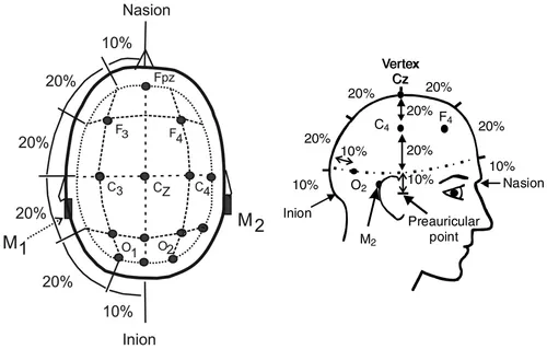
Electroencephalography Monitoring

EEG monitoring to detect and stage sleep requires only a portion of the electrodes used for clinical EEG monitoring. The nomenclature for EEG electrodes follows the International 10-20 system.5 In this system, even-numbered subscripts refer to the right side of the head and odd-numbered subscripts to the left. Electrodes are named for the part of the brain they cover: F for frontal, C for central, and O for occipital (Figure F1-1). The central midline (vertex) electrodes (Cz) and the frontopolar midline electrode (Fpz) are also of interest. The Fpz position is often used for the ground electrode and the Cz position for the reference electrode. Note that before publication of the AASM Scoring Manual, electrodes M1 and M2 were referred to as A1 and A2, respectively. In clinical EEG monitoring, A1 and A2 are, in fact, referred to as earlobe electrodes.
f00-01-9781455770519
Figure F1-1 Nomenclature and position of the basic electrodes for sleep monitoring. C, Central; F, frontal; O, occipital. Even numbers on the right and odd on the left. (Adapted from Berry RB: Fundamentals of sleep medicine, Philadelphia, 2012, Saunders, pp. 2-3.)
The “10-20” in the International 10-20 system of nomenclature for EEG electrodes refers to the fact that the electrodes are positioned at either 10% or 20% of the distance between landmarks.5 The major landmarks include the nasion (bridge of the nose), inion (prominence at base of the occiput), and preauricular points (Figure F1-2).
f00-02-9781455770519
Figure F1-2 Electrode positions using the international 10-20 system. Electrodes are placed at 10% or 20% of the distance between landmarks. (Adapted from Berry RB: Fundamentals of sleep medicine, Philadelphia, 2012, Saunders, pp. 2-3.)

Electroencephalography Derivations

EEG recording uses differential alternating current (AC) amplifiers, which are designed to amplify the difference in voltage between electrodes. There is cancellation of signals common to both electrodes (common mode rejection). (Figure F1-3), this type of AC amplifier allows the recording of relatively low-voltage EEG activity superimposed on a background of high...

Inhaltsverzeichnis