Sleep Medicine Pearls E-Book
eBook - ePub

Sleep Medicine Pearls E-Book

Richard B. Berry, Mary H Wagner

Partager le livre
  1. 580 pages
  2. English
  3. ePUB (adapté aux mobiles)
  4. Disponible sur iOS et Android
eBook - ePub

Sleep Medicine Pearls E-Book

Richard B. Berry, Mary H Wagner

DĂ©tails du livre
Aperçu du livre
Table des matiĂšres
Citations

À propos de ce livre

Sleep Medicine is a rapidly growing and changing field. Experienced sleep medicine clinicians and educators Richard B. Berry, MD and Mary H. Wagner, MD present the completely revised, third edition of Sleep Medicine Pearls featuring 150 cases that review key elements in the evaluation and management of a wide variety of sleep disorders. The cases are preceded by short fundamentals chapters that present enough basic information so that a physician new to sleep medicine can start reading page 1 and quickly learn the essential information needed to care for patients with sleep disorders. A concise, practical format makes this an ideal resource for sleep medicine physicians in active practice, sleep fellows learning sleep medicine, and physicians studying for the sleep boards.

  • Consult this title on your favorite e-reader, conduct rapid searches, and adjust font sizes for optimal readability.
  • Zero in on the practical, "case-based" information you need to effectively interpret sleep studies (polysomnography, home sleep testing, multiple sleep latency testing), sleep logs, and actigraphy.
  • Get clear, visual guidance with numerous figures and sleep tracings illustrating important concepts that teach the reader how to recognize important patterns needed to diagnose sleep disorders.
  • Confer on the go with short, templated chapters—ideal for use by busy physicians. A combination of brief didactic material followed by case-based examples illustrates major points.
  • Stay current with knowledge about the latest developments in sleep medicine by reading updated chapters using the new diagnostic criteria of the recently published International Classification of Sleep Disorder, 3rd Edition and sleep staging and respiratory event scoring using updated versions of the scoring manual of the American Academy of Sleep Medicine Manual for the Scoring of Sleep and Associated Events.
  • Benefit from Drs. Berry and Wagner's 25+ years of clinical experience providing care for patients with sleep disorders and educational expertise from presenting lectures at local, regional and national sleep medicine courses. Dr Berry was awarded the AASM Excellence in Education Award in 2010.

Foire aux questions

Comment puis-je résilier mon abonnement ?
Il vous suffit de vous rendre dans la section compte dans paramĂštres et de cliquer sur « RĂ©silier l’abonnement ». C’est aussi simple que cela ! Une fois que vous aurez rĂ©siliĂ© votre abonnement, il restera actif pour le reste de la pĂ©riode pour laquelle vous avez payĂ©. DĂ©couvrez-en plus ici.
Puis-je / comment puis-je télécharger des livres ?
Pour le moment, tous nos livres en format ePub adaptĂ©s aux mobiles peuvent ĂȘtre tĂ©lĂ©chargĂ©s via l’application. La plupart de nos PDF sont Ă©galement disponibles en tĂ©lĂ©chargement et les autres seront tĂ©lĂ©chargeables trĂšs prochainement. DĂ©couvrez-en plus ici.
Quelle est la différence entre les formules tarifaires ?
Les deux abonnements vous donnent un accĂšs complet Ă  la bibliothĂšque et Ă  toutes les fonctionnalitĂ©s de Perlego. Les seules diffĂ©rences sont les tarifs ainsi que la pĂ©riode d’abonnement : avec l’abonnement annuel, vous Ă©conomiserez environ 30 % par rapport Ă  12 mois d’abonnement mensuel.
Qu’est-ce que Perlego ?
Nous sommes un service d’abonnement Ă  des ouvrages universitaires en ligne, oĂč vous pouvez accĂ©der Ă  toute une bibliothĂšque pour un prix infĂ©rieur Ă  celui d’un seul livre par mois. Avec plus d’un million de livres sur plus de 1 000 sujets, nous avons ce qu’il vous faut ! DĂ©couvrez-en plus ici.
Prenez-vous en charge la synthÚse vocale ?
Recherchez le symbole Écouter sur votre prochain livre pour voir si vous pouvez l’écouter. L’outil Écouter lit le texte Ă  haute voix pour vous, en surlignant le passage qui est en cours de lecture. Vous pouvez le mettre sur pause, l’accĂ©lĂ©rer ou le ralentir. DĂ©couvrez-en plus ici.
Est-ce que Sleep Medicine Pearls E-Book est un PDF/ePUB en ligne ?
Oui, vous pouvez accĂ©der Ă  Sleep Medicine Pearls E-Book par Richard B. Berry, Mary H Wagner en format PDF et/ou ePUB ainsi qu’à d’autres livres populaires dans Medicine et Pulmonary & Thoracic Medicine. Nous disposons de plus d’un million d’ouvrages Ă  dĂ©couvrir dans notre catalogue.

Informations

Éditeur
Saunders
Année
2014
ISBN
9780323319713
Fundamentals 1
Sleep Stage Nomenclature and Basic Monitoring of Sleep

Introduction

Sleep is divided into non–rapid eye movement (NREM) and rapid eye movement (REM) sleep. From 1968 until 2007, sleep was usually staged according to A Manual of Standardized Terminology, Techniques, and Scoring System for Sleep Stages of Human Subjects, edited by Rechtschaffen and Kales (R&K).1 In the R&K Scoring Manual, NREM sleep was divided in stages 1, 2, 3, and 4. REM sleep was referred to as stage REM. Sleep stage nomenclature has changed following the publication of the American Academy of Sleep Medicine (AASM) Manual for the Scoring of Sleep and Associated Events (hereafter referred to as the AASM Scoring Manual) in 2007.2,3 To denote sleep staging by new criteria, sleep stage nomenclature has changed. The old and new nomenclatures are shown in Table F1-1. Stages 3 and stage 4 are combined into stage N3. REM sleep is referred as stage R. An update of the AASM Scoring Manual has recently been published, but sleep stage nomenclature remains unchanged.4
Table F1-1
Sleep Stage Nomenclature
R&KAASM
WakeStage WStage W
NREMStage 1Stage N1
Stage 2Stage N2
Stage 3Stage N3
Stage 4
REMStage REMStage R
AASM, American Academy of Sleep Medicine; NREM, non–rapid eye movement; REM, rapid eye movement; R&K, Rechtschaffen and Kales.
Sleep staging is based on electroencephalography (EEG), electrooculography (EOG), and submental (chin) electromyography (EMG) criteria. EOG (eye movement recording) and chin EMG recordings are used to detect stage R, which is characterized by rapid eye movements (REMs) and reduced muscle tone.

Time Window for Staging Sleep

Digital polysomnography (sleep recording) allows visualization of the waveforms in multiple time windows (10, 15, 30, 60, 90, 120, and 240 seconds) (Table F1-2). A 30-second window is used to stage sleep (known as an epoch), whereas a 10-second window is used for clinical electroencephalography (EEG) monitoring. A 10-second window allows for detailed visualization of waveforms to determine frequency. The convention of using a 30-second window for sleep staging is based on paper recording using ink pens during the early days of sleep monitoring. At a page speed of 10 millimeters per second (mm/s), a standard 30-centimeter (cm) page of recording paper represented 30 seconds. Each page represented one epoch. Sleep is still staged today in sequential 30-second epochs, although digital polysomnography allows for use of different time windows for scoring respiratory and other events.
Table F1-2
Optimal Window Duration for Viewing Events in Polysomnography
Window DurationUse
30 seconds (an epoch)Sleep staging
60–120 secondsRespiratory Events
15 secondsClinical EEG
10 secondsECG rhythms
Identifying wave form frequency
ECG, Electrocardiography; EEG, electroencephalography.

Electroencephalography Monitoring

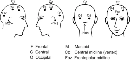
EEG monitoring to detect and stage sleep requires only a portion of the electrodes used for clinical EEG monitoring. The nomenclature for EEG electrodes follows the International 10-20 system.5 In this system, even-numbered subscripts refer to the right side of the head and odd-numbered subscripts to the left. Electrodes are named for the part of the brain they cover: F for frontal, C for central, and O for occipital (Figure F1-1). The central midline (vertex) electrodes (Cz) and the frontopolar midline electrode (Fpz) are also of interest. The Fpz position is often used for the ground electrode and the Cz position for the reference electrode. Note that before publication of the AASM Scoring Manual, electrodes M1 and M2 were referred to as A1 and A2, respectively. In clinical EEG monitoring, A1 and A2 are, in fact, referred to as earlobe electrodes.
f00-01-9781455770519
Figure F1-1 Nomenclature and position of the basic electrodes for sleep monitoring. C, Central; F, frontal; O, occipital. Even numbers on the right and odd on the left. (Adapted from Berry RB: Fundamentals of sleep medicine, Philadelphia, 2012, Saunders, pp. 2-3.)
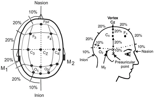
The “10-20” in the International 10-20 system of nomenclature for EEG electrodes refers to the fact that the electrodes are positioned at either 10% or 20% of the distance between landmarks.5 The major landmarks include the nasion (bridge of the nose), inion (prominence at base of the occiput), and preauricular points (Figure F1-2).
f00-02-9781455770519
Figure F1-2 Electrode positions using the international 10-20 system. Electrodes are placed at 10% or 20% of the distance between landmarks. (Adapted from Berry RB: Fundamentals of sleep medicine, Philadelphia, 2012, Saunders, pp. 2-3.)

Electroencephalography Derivations

EEG recording uses differential alternating current (AC) amplifiers, which are designed to amplify the difference in voltage between electrodes. There is cancellation of signals common to both electrodes (common mode rejection). (Figure F1-3), this type of AC amplifier allows the recording of relatively low-voltage EEG activity superimposed on a background of high...

Table des matiĂšres