The Radiology Technologist's Handbook to Surgical Procedures
eBook - ePub

The Radiology Technologist's Handbook to Surgical Procedures

AnthonyC Anderson

Compartir libro
  1. 152 páginas
  2. English
  3. ePUB (apto para móviles)
  4. Disponible en iOS y Android
eBook - ePub

The Radiology Technologist's Handbook to Surgical Procedures

AnthonyC Anderson

Detalles del libro
Vista previa del libro
Índice
Citas

Información del libro

In the past several years, the rapid development of sophisticated imaging modalities has made radiology the fastest growing specialty in medicine. It is important for the radiologic technologist to keep pace with technology's advancements. The influx of freestanding outpatient facilities and the demands of insurance companies, HMOs and third party reimbursement have brought about change. Medical facilities have begun to call upon nurses, surgical technicians, and other non-radiologic personnel to assist with patient positioning during surgical procedures requiring imaging-creating a need for a concise, how-to guide to performing surgical procedures. The Radiology Technologist's Handbook to Surgical Procedures provides a quick reference for using fluoroscopic and x-ray equipment during surgical procedures. This book includes detailed descriptions and photographs taken in actual clinical settings.By using this manual as a foundation, the radiologic technologist will be able to master many of the operating room x-ray procedures.

Preguntas frecuentes

¿Cómo cancelo mi suscripción?
Simplemente, dirígete a la sección ajustes de la cuenta y haz clic en «Cancelar suscripción». Así de sencillo. Después de cancelar tu suscripción, esta permanecerá activa el tiempo restante que hayas pagado. Obtén más información aquí.
¿Cómo descargo los libros?
Por el momento, todos nuestros libros ePub adaptables a dispositivos móviles se pueden descargar a través de la aplicación. La mayor parte de nuestros PDF también se puede descargar y ya estamos trabajando para que el resto también sea descargable. Obtén más información aquí.
¿En qué se diferencian los planes de precios?
Ambos planes te permiten acceder por completo a la biblioteca y a todas las funciones de Perlego. Las únicas diferencias son el precio y el período de suscripción: con el plan anual ahorrarás en torno a un 30 % en comparación con 12 meses de un plan mensual.
¿Qué es Perlego?
Somos un servicio de suscripción de libros de texto en línea que te permite acceder a toda una biblioteca en línea por menos de lo que cuesta un libro al mes. Con más de un millón de libros sobre más de 1000 categorías, ¡tenemos todo lo que necesitas! Obtén más información aquí.
¿Perlego ofrece la función de texto a voz?
Busca el símbolo de lectura en voz alta en tu próximo libro para ver si puedes escucharlo. La herramienta de lectura en voz alta lee el texto en voz alta por ti, resaltando el texto a medida que se lee. Puedes pausarla, acelerarla y ralentizarla. Obtén más información aquí.
¿Es The Radiology Technologist's Handbook to Surgical Procedures un PDF/ePUB en línea?
Sí, puedes acceder a The Radiology Technologist's Handbook to Surgical Procedures de AnthonyC Anderson en formato PDF o ePUB, así como a otros libros populares de Jura y Forensik. Tenemos más de un millón de libros disponibles en nuestro catálogo para que explores.

Información

Editorial
CRC Press
Año
2017
ISBN
9781351409339
Edición
1
Categoría
Jura
Categoría
Forensik
Part I:
Fluoroscopic Examinations
1. FEMORAL NAILING
(AP POSITION)
PATIENT POSITION: Patient will be in lateral position with affected femur up and slightly forward.
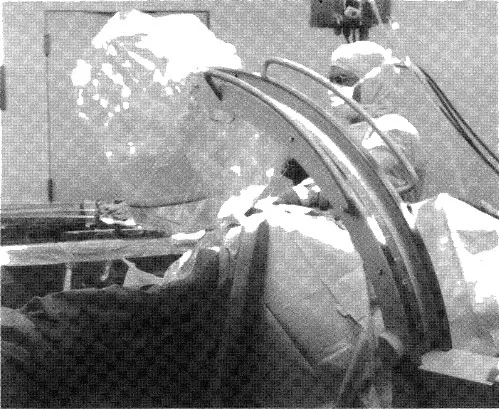
C-ARM POSITION: C-arm will enter facing patient. Rotate c-arm underneath the table to the ap projections. Ensure that c-arm is perpendicular to femur.
Notes: When moving from ap to lateral position, make sure not to bump instrumentation.
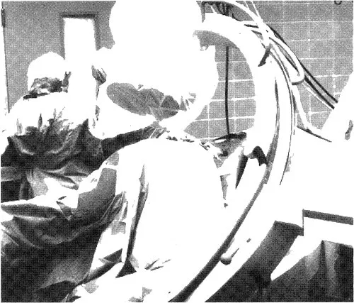
Position c-arm viewing cart at feet of patient with the doctor standing posterior to the patient.
Create distance and magnify image when creating round holes for distal locking screws.
During reaming, obtain true lateral of knee by tilting and rotating c-arm.
Position the c-arm to view reamers crossing fracture site.
Image
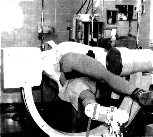
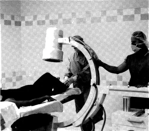
Figure 1.1 C-arm in ap projection of hip.
Image
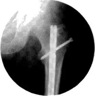
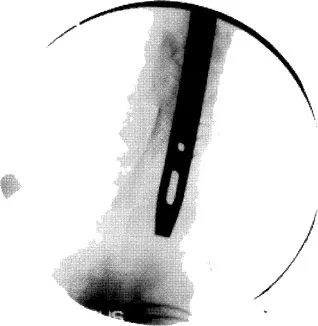
Figure 1.2 C-arm image of ap hip with nail inserted.
2. FEMORAL NAILING
(LATERAL POSITION)
PATIENT POSITION: Patient will be in lateral position with affected femur up and slightly forward.
C-ARM POSITION: C-arm will enter facing patient. Rotate c-arm 10 to 15 degrees over top of patient. Tilt c-arm 5 to 10 degrees toward head of femur.
This view, called the Winquist View, is used to throw the unaffected femur out of view, to elongate the femoral neck for true lateral viewing of starting position and to check anterior and posterior position.
Notes: When moving from ap to lateral position, make sure not to bump instrumentation.
Position c-arm viewing cart at feet of patient.
Doctor will stand posterior to patient.
Image
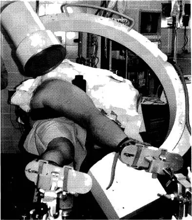
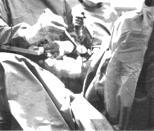
Figure 2.1 C-arm in lateral projection with Winquist tilt view.
Image
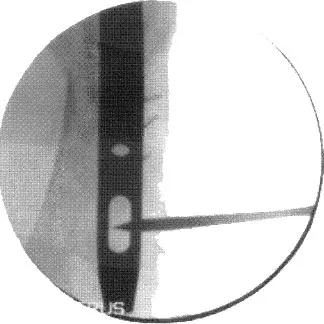
Figure 2.2 C-arm image of lateral hip with starting auld in place.
Image
Figure 2.3 The c-arm is in the Winquist view, which is best utilized to check anterior and posterior positions in relationship to the femoral shaft. The c-arm is rotated over the top and tilted to elongate the femur.
Image
Figure 2.4 The c-arm in a true lateral view in relationship to femur position. The c-arm is tilted to align with the femur. This will give a better indication of fracture alignment and reamer size.
Image
Figure 2.5 During distal targeting for femoral nail, raise the c-arm away from the knee to enlarge the hole and create working space. Use the magnify button on the c-arm if available.
Image
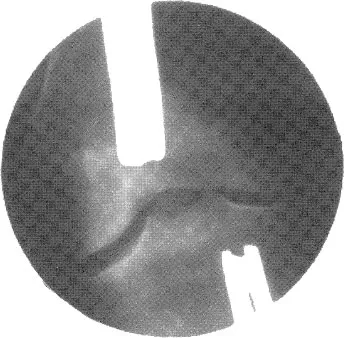
Figure 2.6 Incorrect hole alignment. Note how hole is oblong.
Image
Figure 2.7 Correct hole alignment. Note that hole is now rounded.
3. SUPRACONDYLAR FEMORAL NAIL
(AP VIEW)
PATIENT POSITION: Patient will be supine with the affected knee slightly bent on radiolucent table.
C-ARM POSITION: C-arm will enter perpendicular to patient and in the ap position.
Notes: Ensure underneath clearance allows for movement from the knee to the hip.
C-arm may have to be rotated over or backward to create a true ap view.
Tilt c-arm to open joint space of knee for starting point.
Image
Figure 3.1 C-arm in ap projection of distal femur.
Image
Figure 3.2 X-ray image of distal femur.
2323__...

Índice