The Radiology Technologist's Handbook to Surgical Procedures
eBook - ePub

The Radiology Technologist's Handbook to Surgical Procedures

AnthonyC Anderson

Condividi libro
  1. 152 pagine
  2. English
  3. ePUB (disponibile sull'app)
  4. Disponibile su iOS e Android
eBook - ePub

The Radiology Technologist's Handbook to Surgical Procedures

AnthonyC Anderson

Dettagli del libro
Anteprima del libro
Indice dei contenuti
Citazioni

Informazioni sul libro

In the past several years, the rapid development of sophisticated imaging modalities has made radiology the fastest growing specialty in medicine. It is important for the radiologic technologist to keep pace with technology's advancements. The influx of freestanding outpatient facilities and the demands of insurance companies, HMOs and third party reimbursement have brought about change. Medical facilities have begun to call upon nurses, surgical technicians, and other non-radiologic personnel to assist with patient positioning during surgical procedures requiring imaging-creating a need for a concise, how-to guide to performing surgical procedures. The Radiology Technologist's Handbook to Surgical Procedures provides a quick reference for using fluoroscopic and x-ray equipment during surgical procedures. This book includes detailed descriptions and photographs taken in actual clinical settings.By using this manual as a foundation, the radiologic technologist will be able to master many of the operating room x-ray procedures.

Domande frequenti

Come faccio ad annullare l'abbonamento?
È semplicissimo: basta accedere alla sezione Account nelle Impostazioni e cliccare su "Annulla abbonamento". Dopo la cancellazione, l'abbonamento rimarrà attivo per il periodo rimanente già pagato. Per maggiori informazioni, clicca qui
È possibile scaricare libri? Se sì, come?
Al momento è possibile scaricare tramite l'app tutti i nostri libri ePub mobile-friendly. Anche la maggior parte dei nostri PDF è scaricabile e stiamo lavorando per rendere disponibile quanto prima il download di tutti gli altri file. Per maggiori informazioni, clicca qui
Che differenza c'è tra i piani?
Entrambi i piani ti danno accesso illimitato alla libreria e a tutte le funzionalità di Perlego. Le uniche differenze sono il prezzo e il periodo di abbonamento: con il piano annuale risparmierai circa il 30% rispetto a 12 rate con quello mensile.
Cos'è Perlego?
Perlego è un servizio di abbonamento a testi accademici, che ti permette di accedere a un'intera libreria online a un prezzo inferiore rispetto a quello che pagheresti per acquistare un singolo libro al mese. Con oltre 1 milione di testi suddivisi in più di 1.000 categorie, troverai sicuramente ciò che fa per te! Per maggiori informazioni, clicca qui.
Perlego supporta la sintesi vocale?
Cerca l'icona Sintesi vocale nel prossimo libro che leggerai per verificare se è possibile riprodurre l'audio. Questo strumento permette di leggere il testo a voce alta, evidenziandolo man mano che la lettura procede. Puoi aumentare o diminuire la velocità della sintesi vocale, oppure sospendere la riproduzione. Per maggiori informazioni, clicca qui.
The Radiology Technologist's Handbook to Surgical Procedures è disponibile online in formato PDF/ePub?
Sì, puoi accedere a The Radiology Technologist's Handbook to Surgical Procedures di AnthonyC Anderson in formato PDF e/o ePub, così come ad altri libri molto apprezzati nelle sezioni relative a Jura e Forensik. Scopri oltre 1 milione di libri disponibili nel nostro catalogo.

Informazioni

Editore
CRC Press
Anno
2017
ISBN
9781351409339
Edizione
1
Argomento
Jura
Categoria
Forensik
Part I:
Fluoroscopic Examinations
1. FEMORAL NAILING
(AP POSITION)
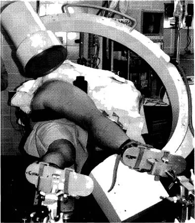
PATIENT POSITION: Patient will be in lateral position with affected femur up and slightly forward.
C-ARM POSITION: C-arm will enter facing patient. Rotate c-arm underneath the table to the ap projections. Ensure that c-arm is perpendicular to femur.
Notes: When moving from ap to lateral position, make sure not to bump instrumentation.
Position c-arm viewing cart at feet of patient with the doctor standing posterior to the patient.
Create distance and magnify image when creating round holes for distal locking screws.
During reaming, obtain true lateral of knee by tilting and rotating c-arm.
Position the c-arm to view reamers crossing fracture site.
Image
Figure 1.1 C-arm in ap projection of hip.
Image
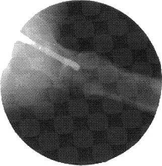
Figure 1.2 C-arm image of ap hip with nail inserted.
2. FEMORAL NAILING
(LATERAL POSITION)
PATIENT POSITION: Patient will be in lateral position with affected femur up and slightly forward.
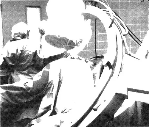
C-ARM POSITION: C-arm will enter facing patient. Rotate c-arm 10 to 15 degrees over top of patient. Tilt c-arm 5 to 10 degrees toward head of femur.
This view, called the Winquist View, is used to throw the unaffected femur out of view, to elongate the femoral neck for true lateral viewing of starting position and to check anterior and posterior position.
Notes: When moving from ap to lateral position, make sure not to bump instrumentation.
Position c-arm viewing cart at feet of patient.
Doctor will stand posterior to patient.
Image
Figure 2.1 C-arm in lateral projection with Winquist tilt view.
Image
Figure 2.2 C-arm image of lateral hip with starting auld in place.
Image
Figure 2.3 The c-arm is in the Winquist view, which is best utilized to check anterior and posterior positions in relationship to the femoral shaft. The c-arm is rotated over the top and tilted to elongate the femur.
Image
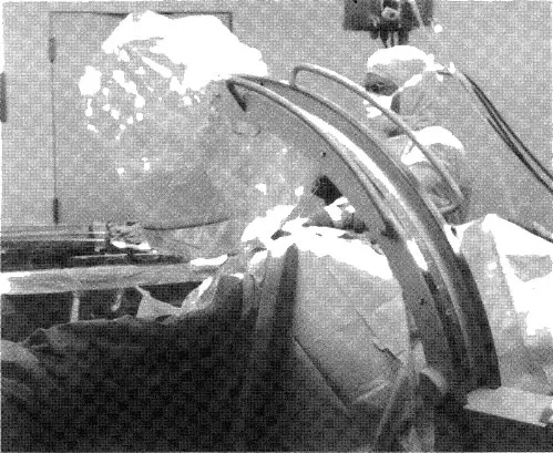
Figure 2.4 The c-arm in a true lateral view in relationship to femur position. The c-arm is tilted to align with the femur. This will give a better indication of fracture alignment and reamer size.
Image
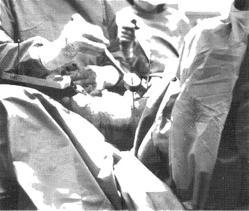
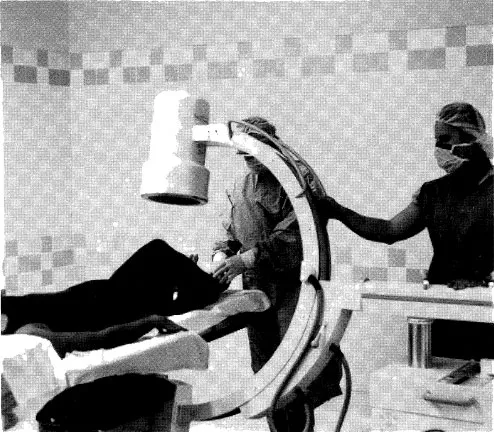
Figure 2.5 During distal targeting for femoral nail, raise the c-arm away from the knee to enlarge the hole and create working space. Use the magnify button on the c-arm if available.
Image
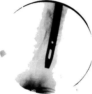
Figure 2.6 Incorrect hole alignment. Note how hole is oblong.
Image
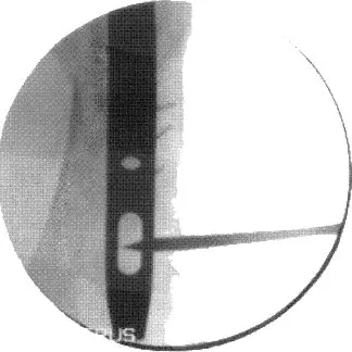
Figure 2.7 Correct hole alignment. Note that hole is now rounded.
3. SUPRACONDYLAR FEMORAL NAIL
(AP VIEW)
PATIENT POSITION: Patient will be supine with the affected knee slightly bent on radiolucent table.
C-ARM POSITION: C-arm will enter perpendicular to patient and in the ap position.
Notes: Ensure underneath clearance allows for movement from the knee to the hip.
C-arm may have to be rotated over or backward to create a true ap view.
Tilt c-arm to open joint space of knee for starting point.
Image
Figure 3.1 C-arm in ap projection of distal femur.
Image
Figure 3.2 X-ray image of distal femur.
2323__...

Indice dei contenuti