The Radiology Technologist's Handbook to Surgical Procedures
eBook - ePub

The Radiology Technologist's Handbook to Surgical Procedures

AnthonyC Anderson

Buch teilen
  1. 152 Seiten
  2. English
  3. ePUB (handyfreundlich)
  4. Über iOS und Android verfügbar
eBook - ePub

The Radiology Technologist's Handbook to Surgical Procedures

AnthonyC Anderson

Angaben zum Buch
Buchvorschau
Inhaltsverzeichnis
Quellenangaben

Über dieses Buch

In the past several years, the rapid development of sophisticated imaging modalities has made radiology the fastest growing specialty in medicine. It is important for the radiologic technologist to keep pace with technology's advancements. The influx of freestanding outpatient facilities and the demands of insurance companies, HMOs and third party reimbursement have brought about change. Medical facilities have begun to call upon nurses, surgical technicians, and other non-radiologic personnel to assist with patient positioning during surgical procedures requiring imaging-creating a need for a concise, how-to guide to performing surgical procedures. The Radiology Technologist's Handbook to Surgical Procedures provides a quick reference for using fluoroscopic and x-ray equipment during surgical procedures. This book includes detailed descriptions and photographs taken in actual clinical settings.By using this manual as a foundation, the radiologic technologist will be able to master many of the operating room x-ray procedures.

Häufig gestellte Fragen

Wie kann ich mein Abo kündigen?
Gehe einfach zum Kontobereich in den Einstellungen und klicke auf „Abo kündigen“ – ganz einfach. Nachdem du gekündigt hast, bleibt deine Mitgliedschaft für den verbleibenden Abozeitraum, den du bereits bezahlt hast, aktiv. Mehr Informationen hier.
(Wie) Kann ich Bücher herunterladen?
Derzeit stehen all unsere auf Mobilgeräte reagierenden ePub-Bücher zum Download über die App zur Verfügung. Die meisten unserer PDFs stehen ebenfalls zum Download bereit; wir arbeiten daran, auch die übrigen PDFs zum Download anzubieten, bei denen dies aktuell noch nicht möglich ist. Weitere Informationen hier.
Welcher Unterschied besteht bei den Preisen zwischen den Aboplänen?
Mit beiden Aboplänen erhältst du vollen Zugang zur Bibliothek und allen Funktionen von Perlego. Die einzigen Unterschiede bestehen im Preis und dem Abozeitraum: Mit dem Jahresabo sparst du auf 12 Monate gerechnet im Vergleich zum Monatsabo rund 30 %.
Was ist Perlego?
Wir sind ein Online-Abodienst für Lehrbücher, bei dem du für weniger als den Preis eines einzelnen Buches pro Monat Zugang zu einer ganzen Online-Bibliothek erhältst. Mit über 1 Million Büchern zu über 1.000 verschiedenen Themen haben wir bestimmt alles, was du brauchst! Weitere Informationen hier.
Unterstützt Perlego Text-zu-Sprache?
Achte auf das Symbol zum Vorlesen in deinem nächsten Buch, um zu sehen, ob du es dir auch anhören kannst. Bei diesem Tool wird dir Text laut vorgelesen, wobei der Text beim Vorlesen auch grafisch hervorgehoben wird. Du kannst das Vorlesen jederzeit anhalten, beschleunigen und verlangsamen. Weitere Informationen hier.
Ist The Radiology Technologist's Handbook to Surgical Procedures als Online-PDF/ePub verfügbar?
Ja, du hast Zugang zu The Radiology Technologist's Handbook to Surgical Procedures von AnthonyC Anderson im PDF- und/oder ePub-Format sowie zu anderen beliebten Büchern aus Jura & Forensik. Aus unserem Katalog stehen dir über 1 Million Bücher zur Verfügung.

Information

Verlag
CRC Press
Jahr
2017
ISBN
9781351409339
Auflage
1
Thema
Jura
Part I:
Fluoroscopic Examinations
1. FEMORAL NAILING
(AP POSITION)
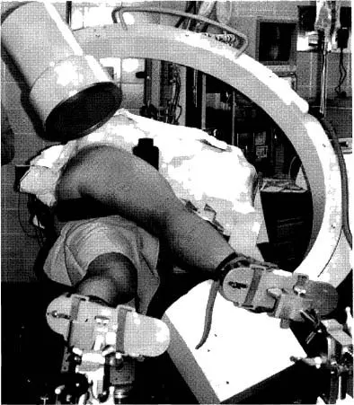
PATIENT POSITION: Patient will be in lateral position with affected femur up and slightly forward.
C-ARM POSITION: C-arm will enter facing patient. Rotate c-arm underneath the table to the ap projections. Ensure that c-arm is perpendicular to femur.
Notes: When moving from ap to lateral position, make sure not to bump instrumentation.
Position c-arm viewing cart at feet of patient with the doctor standing posterior to the patient.
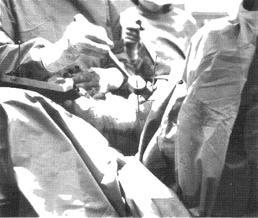
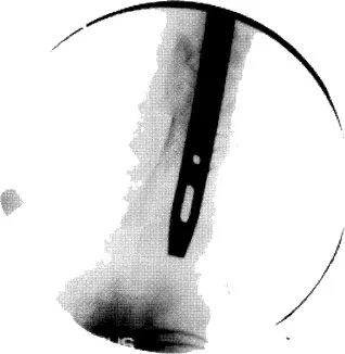
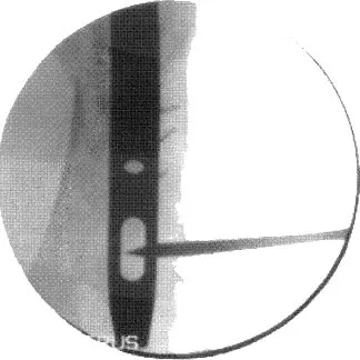
Create distance and magnify image when creating round holes for distal locking screws.
During reaming, obtain true lateral of knee by tilting and rotating c-arm.
Position the c-arm to view reamers crossing fracture site.
Image
Figure 1.1 C-arm in ap projection of hip.
Image
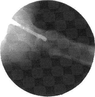
Figure 1.2 C-arm image of ap hip with nail inserted.
2. FEMORAL NAILING
(LATERAL POSITION)
PATIENT POSITION: Patient will be in lateral position with affected femur up and slightly forward.
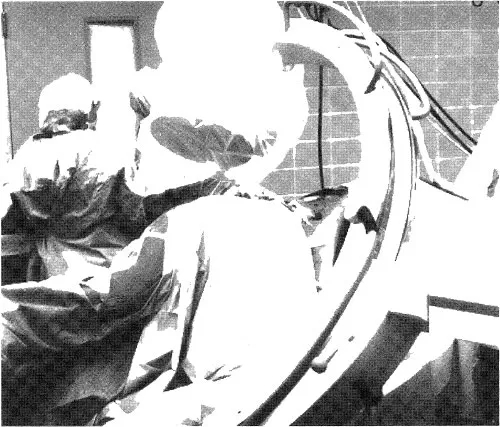
C-ARM POSITION: C-arm will enter facing patient. Rotate c-arm 10 to 15 degrees over top of patient. Tilt c-arm 5 to 10 degrees toward head of femur.
This view, called the Winquist View, is used to throw the unaffected femur out of view, to elongate the femoral neck for true lateral viewing of starting position and to check anterior and posterior position.
Notes: When moving from ap to lateral position, make sure not to bump instrumentation.
Position c-arm viewing cart at feet of patient.
Doctor will stand posterior to patient.
Image
Figure 2.1 C-arm in lateral projection with Winquist tilt view.
Image
Figure 2.2 C-arm image of lateral hip with starting auld in place.
Image
Figure 2.3 The c-arm is in the Winquist view, which is best utilized to check anterior and posterior positions in relationship to the femoral shaft. The c-arm is rotated over the top and tilted to elongate the femur.
Image
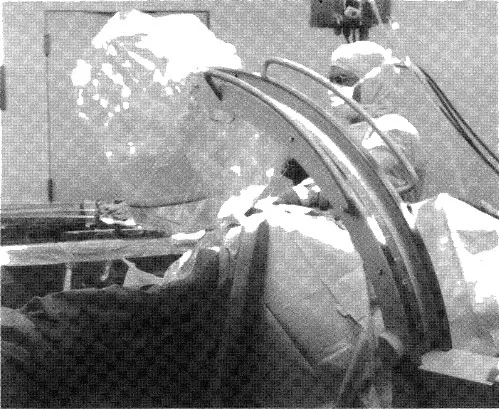
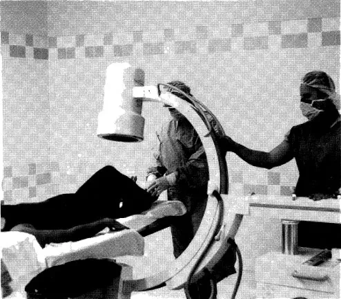
Figure 2.4 The c-arm in a true lateral view in relationship to femur position. The c-arm is tilted to align with the femur. This will give a better indication of fracture alignment and reamer size.
Image
Figure 2.5 During distal targeting for femoral nail, raise the c-arm away from the knee to enlarge the hole and create working space. Use the magnify button on the c-arm if available.
Image
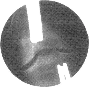
Figure 2.6 Incorrect hole alignment. Note how hole is oblong.
Image
Figure 2.7 Correct hole alignment. Note that hole is now rounded.
3. SUPRACONDYLAR FEMORAL NAIL
(AP VIEW)
PATIENT POSITION: Patient will be supine with the affected knee slightly bent on radiolucent table.
C-ARM POSITION: C-arm will enter perpendicular to patient and in the ap position.
Notes: Ensure underneath clearance allows for movement from the knee to the hip.
C-arm may have to be rotated over or backward to create a true ap view.
Tilt c-arm to open joint space of knee for starting point.
Image
Figure 3.1 C-arm in ap projection of distal femur.
Image
Figure 3.2 X-ray image of distal femur.
2323__...

Inhaltsverzeichnis