Biological Sciences
Cell Wall
The cell wall is a rigid, protective layer that surrounds the cell membrane of plant cells, fungi, and some bacteria. It provides structural support and protection, helping the cell maintain its shape and resist mechanical stress. Composed mainly of cellulose in plants, the cell wall also regulates the passage of molecules in and out of the cell.
Written by Perlego with AI-assistance
Related key terms
1 of 5
12 Key excerpts on "Cell Wall"
 eBook - PDF
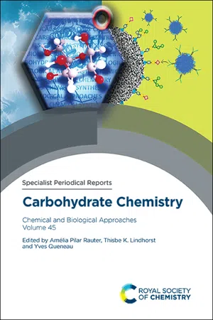
Carbohydrate Chemistry
Chemical and Biological Approaches Volume 45
- Amélia Pilar Rauter, Thisbe K Lindhorst, Yves Queneau, Amélia Pilar Rauter, Thisbe K Lindhorst, Yves Queneau(Authors)
- 2021(Publication Date)
- Royal Society of Chemistry(Publisher)
The plant Cell Wall is a complex structure that must satisfy multiple functions: it must exhibit a sufficient tensile strength to support internal turgor pressure and must provide enough flexibility to allow cell division and elongation. The plant Cell Wall also forms a physical barrier that protects the protoplast against biotic and abiotic stresses. Plant Cell Walls are responsible for shaping the different cell types found in plant tissues and organs and also play roles in inter-cellular communication. All plant Cell Walls are composed of two layers; the middle lamella and the primary Cell Wall. The middle lamella provides the adhesion between adjacent cells, whereas the primary wall is built up by each individual cell during division and expansion. When cell growth is achieved, the plant cell maintains its primary Cell Wall but may also deposit a secondary Cell Wall. During the synthesis of this secondary Cell Wall, cellulose, hemi-cellulose and lignin are deposited to build fibres and vascular bundles for water and mineral transport and to ensure most of the mechanical support of the plant body. Going back to the primary Cell Wall, its main a Normandie Univ, UNIROUEN, SFR NORVEGE, Innovation Chimie Carnot, Laboratoire Glycobiologie et Matrice Extracellulaire ve ´ge ´tale EA4358, 76000 Rouen, France. E-mail: [email protected] b Normandie Univ, UNIROUEN, INSA Rouen, CNRS, Innovation Chimie Carnot, COBRA (UMR6014), 76000 Rouen, France Carbohydr. Chem. , 2021, 45 , 553–571 | 553 c The Royal Society of Chemistry 2022 components are cellulose, hemicelluloses and pectic polysaccharides together with some proteins and phenolic compounds. Together, poly-saccharides represent more than 90% of the primary Cell Wall biomass. Pectins are acidic polysaccharides that predominantly exist in the pri-mary Cell Wall and play crucial roles in cell expansion and strength. 1 They are the most complex and heterogeneous polysaccharides of the primary Cell Wall. eBook - ePub
- Kirsi-Marja Oksman-Caldentey, Wolfgang H. Barz, Kirsi-Marja Oksman-Caldentey, Wolfgang H. Barz(Authors)
- 2002(Publication Date)
- CRC Press(Publisher)
It was concluded that the Cell Wall must have important functional roles, e.g., in cell-cell communication, transport of metabolites, differentiation, and development. To indicate this new appreciation of the Cell Wall as a dynamic and regulatory extracellular organelle, some authors prefer to call it an extracellular matrix. The growing realization that this matrix forms an integral and essential part of the plant cell may eventually even lead us to abandon the term extracellular in favor of, e.g., pericellular. In spite of these considerations, plant Cell Walls do play important static structural roles. First, they have to bear the turgor pressure, which results in enormous tangentially oriented stress forces in the Cell Wall plane (2). This stress is taken up by the fibrillar component in the Cell Wall, namely the cellulose microfibrils, which are interconnected and held in place by hemicelluloses. Second, the Cell Wall has to take up the pressure exerted on a plant cell by the surrounding tissue. This pressure is taken up by the matrix in which the fibrils are embedded, made up of polysaccharides containing uronic acids, namely pectins and glucuronoarabinoxylans, or—more precisely—by the water molecules held in the Cell Walls by these negatively charged polymers. In extreme cases, this water is replaced by the incorporation of lignin, a heavily cross-linked three-dimensional polyphenolic network able to withstand very high pressures. FIGURE 1 In vivo roles and ex vivo uses of plant Cell Walls and their components. Cell Walls fulfill many roles—both structural and functional— in the life of a plant, and different components are responsible for the different roles. Biotechnology may aim at improving these in vivo roles according to human interest, e.g., increasing resistance of crop plants against pathogens (HR, hypersensitive reaction) eBook - ePub
- William V. Dashek, Gurbachan S. Miglani, William V. Dashek, Gurbachan S. Miglani(Authors)
- 2016(Publication Date)
- Wiley(Publisher)
CHAPTER 9 Plant Cell WallsJames E. Bidlack1 and William V. Dashek21Department of Biology, University of Central Oklahoma, Edmond, OK, USA2Retired Faculty, Adult Degree Program, Mary Baldwin College, Staunton, VA, USAIntroduction
Constituents of the plant Cell Wall (CW) are among the most abundant biological molecules on earth and the functions of the CW are diverse. The CW provides mechanical strength, maintains cell shape, controls cell expansion, regulates transport, provides protection, functions in signaling processes, and stores food reserves (Brett and Waldron, 1990). The CW has even been referred to as a cell organelle (Mauseth, 1988), and more information is needed to have a complete understanding of how CW structure and function are integrated.Structure
There are two types of CWs: primary and secondary (Hayashi, 2006; Albersheim et al., 2010). Primary CWs (Figure 9.1 a and b) are thin, elastic, and permit cell expansion while providing strength (Rose, 2003). Cells that possess only primary walls can lose their specialized form, divide, and subsequently differentiate into new cells (Evert, 2006). In some instances, cells possessing primary walls can exhibit thickenings and layering, for example, certain collenchyma cells (Bidlack and Jansky, 2014). The middle lamella is an area of union between adjacent cells’ primary walls. Secondary walls are thick, often consisting of layers and deposited when cell expansion is complete (Carpita et al., 2001). Examples of cells possessing secondary walls are xylem fibers, tracheids, vessel elements, and sclereids (see Chapter 1 ). These cell types possess lignified, spiral, scalariform, reticulate, or pitted walls (Figure 9.2 ).From: Courtesy of J. Mayfield. (b) Model of the primary Cell Wall.(a) Electron micrograph of two adjoining plant cells with an intervening primary wall.Figure 9.1From: https://en.wikipedia.org/wiki/Cell_wall .Figure 9.2 eBook - PDF
- Gurnah, Akinloye(Authors)
- 2018(Publication Date)
- Agri Horti Press(Publisher)
Such walls are relatively impermeable to water. Hemicell-uloses are a poorly defined group of poly saccharides associated with cellulose in plant Cell Walls. They are not chemically related to cellulose, as the name implies, but possess very different chemical and physical properties. Callose is the name given to a carbohydrate membrane substance found in the perforated septa (sieve plates) of the sieve tubes. Similar material presumably of the same chemical composition, has been found in pollen grains and constitutes the inner layer of pollen tubes. It has also been reported as occurring in the fungi. The exact chemical composition of callose is unknown since it has never been obtained in sufficient amounts to permit quantitative determinations. Chitin, a nitrogenous substance common in the exoskeleton of insects, is a constituent of the walls of many fungi and bacteria. It has been reported as being present in the walls of certain algae but it is unknown in any of the higher plants. Tannins are commonly found in the cell sap but they also occur in the walls of certain tissues, especially cork and wood cells. Mucilages are common constituents of the outer walls of many water plants and occur also in the outer walls of some seed coats, in glandular hairs and in other specialized tissues. Inorganic compounds such as silica and salts of calcium, iron, and other metals are also present in some plant Cell Walls. None of these inorganic compounds, however, are regarded as essential constituents of the Cell Wall. This ebook is exclusively for this university only. Cannot be resold/distributed. Plant Cell and their Functions 63 Protoplasm In most mature cells of the vascular plants protoplasmis present only as a thin layer covering the inner surface of the Cell Walls but in some specialized cells branching strands of protoplasm also extend across the vacuole. eBook - ePub
- Charles E. Wyman(Author)
- 2013(Publication Date)
- Wiley(Publisher)
The thickness of the walls in xylem and fiber tissues is due to the formation of an extensive secondary Cell Wall as the cell matures. The sequence of events in the development of these cells is that after the cells have ceased growing by elongation, they begin to deposit a multilamellar secondary Cell Wall toward the cell lumen side of the primary Cell Wall [16]. At some point during and following secondary wall formation, the entire wall is infused with lignin monolignols [17]. The lignin polymerizes into the spaces in the existing wall and, consequently, significantly lowers its porosity [18]. Finally, the cell dies and its contents are absorbed, leaving a rigid water-impermeable cell-wall barrier. The biomass conversion perspective of the plant Cell Wall is, by necessity, heavily skewed toward the thick, lignified, secondary Cell Walls. These are the walls that harbor most of the mass and therefore most of the structural sugars in biomass. Unfortunately, they are also the most recalcitrant.Chemically, the plant Cell Wall is understood to be composed of cellulose, hemicelluloses, pectins, proteins, and often lignin. However, it is the complex intermingling of cross-linked layers brought together largely by self-assembly and template-assembly processes that create the complexity and resilience of the plant Cell Wall. The complex macromolecular architecture of the Cell Wall is a result of the self-ordering properties of cell-wall polymers generating a high degree of structural organization and complexity [19]. Three themes that govern the architectural plan of the plant Cell Wall are: (1) fibrous structural units embedded in an amorphous matrix; (2) covalent and non-covalent cross-linking; and (3) a polylamellate construction [20].The plant Cell Wall is sometimes envisioned as three intermingled networks that are extensively restructured during development. One is a protein network formed by the various classes of glycoproteins that contribute a scaffold to initial cell plate [21]. This network organizes cell plate membranes and the incorporation of initial cell-wall components as they are delivered to the cell plate by Golgi-derived vesicles. This original scaffold gets extensively remodeled and recycled during cell growth and maturation. While the protein network is not usually considered a significant contributor to recalcitrance, its role in establishing a template for cell-wall construction is still important. The second network is the pectic polysaccharide network. In the primary Cell Wall, the pectin network is credited with dictating key structural and mechanical properties of the wall including water content and porosity [22]. Again, this network is remodeled during growth and development and is usually not of critical concern for biomass conversion because the pectic polysaccharides are not especially recalcitrant to hydrolysis and extraction by pretreatment. eBook - PDF
- Jocelyn K. C. Rose(Author)
- 2009(Publication Date)
- Wiley-Blackwell(Publisher)
However, for the purposes of the analytical chemist the wall is the insoluble material that remains after plant tissue or cells have been lysed and then treated with aqueous buffers, organic solvents and enzymes. This isolated wall contains much of the apoplastic content of the tissue but may also contain some cytoplasmic and vacuolar material. Some of the apoplastic material is inevitably lost during the isola-tion of walls even though it may be a component of the wall in vivo . Several investigators have proposed that the terms ‘extracellular matrix’ (Rob-erts, 1989) or ‘exocellular matrix’ (Wyatt and Carpita, 1993) are more appropriate than ‘Cell Wall’ because they suggest a dynamic organelle rather than an inert rigid box. These new terms were not met with universal approval partly because plant scientists have yet to agree on the relationship between a plant cell and its ‘wall’ (Staehelin, 1991). Nevertheless, this debate did serve to draw the attention of a much wider audience to the biological significance of the ‘wall’. Most, if not all, plant sci-entists now agree that ‘… walls do not a prison make…’ (Roberts, 1994) even though they still ‘ call a wall a wall ’. COMPOSITION AND STRUCTURE 3 1.3 The composition of the primary Cell Wall Primary walls isolated from higher plant tissues and cells are composed predomi-nantly of polysaccharides (up to 90% of the dry weight) together with lesser amounts of structural glycoproteins (2–10%), phenolic esters (<2%), ionically and covalently bound minerals (1–5%), and enzymes. Lignin is a characteristic component of sec-ondary walls and is discussed in Chapter 5 of this book. In living tissue water may account for up to 70% of the volume of a primary wall (Monro et al ., 1976). Twelve different glycosyl residues (Figure 1.1) have been shown to be constituents of all primary walls, albeit in different amounts. eBook - ePub
- Malgorzata Lekka, Daniel Navajas, Manfred Radmacher, Alessandro Podestà, Malgorzata Lekka, Daniel Navajas, Manfred Radmacher, Alessandro Podestà(Authors)
- 2023(Publication Date)
- De Gruyter(Publisher)
Overvoorde et al., 2010 , Somssich et al., 2016 ).4.4.2 Plant Cell Walls
Plant Cell Walls are extracellular matrices made of interacting networks of biopolymers and are categorized into primary walls, which are laid down before and during cell expansion, and secondary walls, which are deposited mainly after growth has ceased. The primary Cell Wall is composed of three major classes of polysaccharides: cellulose, pectins, and hemicelluloses, plus smaller amounts of structural proteins, enzymes, small molecules, and water. Together, these components act centrally in the coordinated growth of plant cells. The main driving force of growth comes from turgor pressure that accumulates within the plant cell. The internal environment of a plant cell often has a larger negative osmotic potential due to high solute concentrations within the cell. This promotes transport of water into the vacuole of the cell, increasing turgor pressure. Without the presence of the Cell Wall, the cell would expand in response to this increase in pressure until it reached the maximum volume allowed by the plasma membrane and then burst. To prevent this, the Cell Wall must have high tensile strength to withstand the turgor pressure generated in the cell. However, if the primary Cell Wall were merely a rigid cage that stopped cell expansion, then the plant would never be able to grow. This is where the Cell Wall’s second major physical attribute comes in to play. A high-enough turgor pressure within the cell can cause cellulose microfibrils to slip past each other, allowing the wall to yield and the cell to expand irreversibly (Cosgrove, 2018 ). Not all deformations are irreversible in Cell Walls. Cross-linking of matrix polysaccharides, pectins and hemicelluloses, are vital to the elasticity of the Cell Wall, allowing it to expand temporarily and then return to its previous state (Abasolo et al., 2009 ). This gives plants the flexibility to bend without breaking, a vital trait for organisms that cannot move to take shelter from wind and rain, for example. In primary walls, hemicelluloses (primarily xyloglucan in eudicots or xylans in grasses) and pectins (primarily homogalacturonan (HG)) interact closely with cellulose to maintain the integrity of the Cell Wall. The points at which Cell Wall components come together are called biomechanical hotspots, from which the majority of the strength of the wall arises (Cosgrove, 2018 eBook - PDF
Cells and Their Component Parts
Biochemistry, Physiology, Morphology
- Jean Brachet, Alfred E. Mirsky(Authors)
- 2014(Publication Date)
- Academic Press(Publisher)
CHAPTER 2 Plant Cell Walls By KURT MÜHLETHALER I. The Chemical Nature and Submicroscopic Structure of the Constituents of the Cell Wall 86 A. Cellulose 86 B. Hemicelluloses 88 C. Pectic Substances 88 D. Lignin 89 E. Cuticular Substances 90 F. Mineral Deposits 90 II. Microscopic Structure of Cell Walls 91 III. Indirect Methods of Investigation 94 A. Swelling Characteristics 94 B. Polarized Light Microscopy 95 C. X-Ray Diffraction Methods 98 IV. The Cell Wall in the Electron Microscope 99 A. The Structure of Microfibrils 99 B. Structure of the Primary Wall 101 C. Secondary Cell Wall 104 D. The Tertiary Cell Wall 106 V. The Mechanism of Orientation and Growth 107 A. General 107 B. Tearing Growth 110 C. Multinet Growth 112 D. Tip Growth 113 E. Mosaic Growth 113 F. Extracellular Wall Formation 114 VI. The Structure of Cell Connections 120 VII. Membrane Structure in Various Cell Types 124 A. Fibers and Tracheids 124 B. Collenchyma Cells 125 C. Water-conducting Elements 126 D. Epidermal Cells 127 E. Pollen-Grain Cell Walls 129 VIII. Summary 131 References 131 85 86 KURT MUHLETHALER I. THE CHEMICAL NATURE AND SUBMICROSCOPIC STRUCTURE OF THE CONSTITUENTS OF THE Cell Wall A. Cellulose The most important constituent of the plant Cell Wall is cellulose. It forms the structural framework within which other wall substances, such as pectin, lignin, hemicellulose, etc., are embedded. For this reason, as well as its technical importance, cellulose has been intensively inves-tigated, and many of its chemical, physical, and morphological prop-erties are known. For more than a century, it has been known that this substance hydrolyzes in strong acid to give glucose, and, in fact, the yield is almost quantitative. The elementary structural unit, glucose, has been shown by Haworth (1925) to be a six-membered ring structure with an oxygen bridge connecting carbon atoms numbers 1 and 5. eBook - PDF
- Emea, A(Authors)
- 2018(Publication Date)
- Agri Horti Press(Publisher)
The wall of cells consists of two major parts : 1. The middle lamella and 2. The primary wall. Fig. Perspective View of a Palisade Cell from the Leaf Mesophyll. In walls of many plant cells a third structural component, the secondary wall, is also present. Although some plant cells are known which do not have a well defined Cell Wall, this structure is so generally present in plant cells as to be considered one of their characteristic features. Lining the interior of the wall and occupying more or less of the cell cavity-nucleus is the protoplasm. The protoplasm of active cells is a transparent, slightly viscous, granular material that any conspicuous structural background. It is not homoge-neous, however, and vacucle contains a number of definite structures. One of these, the nucleus, is a denser body which is more or less pheroidal in shape and is separated from the remaining protoplasm by a definite membrane, the nuclear membrane. Perspective view of a rounding the nucleus are : • A clear palisade cell from the leaf mesophyll. liquid known as the nuclear sap, This ebook is exclusively for this university only. Cannot be resold/distributed. The Structure of Plant Cell Wall 85 • A delicate network of denser material, the reticulum, and • One or more small spherical masses of material known as the nucleolus or nucleoli. All of the protoplasm outside of the nucleus of the cell constitutes the cytoplasm. In a typical mature plant cell the cytoplasm is present as a thin layer lining the inner surface of the Cell Wall. The two boundary layers ofthe cytoplasm that in contact with the Cell Wall and that in contact with vacuole are called the cytoplasmic membranes. Imbedded in the cytoplasm are numerous well differentiated bodies known as plastids. Plastids are specialized cytoplasmic structures which are usually centres of certain types of physiological activity. eBook - ePub
- Caroline Bowsher, Alyson Tobin(Authors)
- 2021(Publication Date)
- Garland Science(Publisher)
3 Plant Cell Structure Key Concepts The internal organization of plant cells is dependent on membranes. Membranes, which consist of lipids and proteins, depend on a water phase for their organization into two-dimensional sheets. Proteins are targeted to specific membranes and confer specific properties on the membrane, including its permeability to solutes. The plasma membrane defines the outer boundary of the cell, regulates solute exchange, and participates in Cell Wall formation. Vacuoles can occupy a very large proportion of cell space in vegetative tissues and store a variety of compounds, including pigments, proteins, and osmotically active solutes. The endomembrane system consists of two distinct compartments: the endoplasmic reticulum and the Golgi. It provides a complex internal space bounded by a membrane that is involved in the synthesis of proteins, polysaccharides, and lipids, mostly destined for export from the cell. Cell Walls act as mechanical supports and osmoregulators and define the shape of each cell, ultimately defining the shape of the whole plant. Cell Walls regulate the flux of molecules between cells and provide transport pathways within the plant. In common with other eukaryotic cells, most plant cells contain nuclei, which are concerned with the storage and expression of genetic information; mitochondria, which are mostly involved in respiration; and peroxisomes, which have diverse roles in plants. Plastids are a defining feature of plant cells and are specialized for a variety of functions, notably photosynthesis in all green tissues. Plant Organs and Tissues Consist of Communities of Cells The previous chapter (Chapter 2) emphasized the importance of plant cell structure in the biochemical functioning of the whole plant. It is the unique challenges plants face as sessile organisms that need to, for example, harvest solar energy, acquire inorganic mineral elements, cope with loss of water, etc., that determine their cell structure eBook - ePub
Annual Plant Reviews, Plant Polysaccharides
Biosynthesis and Bioengineering
- Peter Ulvskov(Author)
- 2010(Publication Date)
- Wiley-Blackwell(Publisher)
We are on the verge of a rapid expansion in our understanding of the cell biology of the complex sets of polysaccharides that comprise plant Cell Walls. As we develop tools to map Cell Wall microheterogeneity we can perceive their considerable molecular complexity and diversity. Current major gaps in our knowledge of Cell Walls in relation to growth and development include systematic knowledge of the precise configurations and interactions of polysaccharides within the diverse Cell Walls of a growing organ, the functions of individual polysaccharides within a specific wall composites and also how the structural variants of the polysaccharide classes variedly influence Cell Wall architectures and properties. Inherent in all these studies will be the elucidation of regulatory and controlling mechanisms supporting the assembly of functionally specific wall architectures. At the level of organs, major gaps in knowledge include how cell and tissue wall architectures are integrated into the generation of organ mechanical properties and the nature of the involvement of Cell Walls in responses to diverse environmental and mechanical impacts.AcknowledgementsJPK acknowledges funding support from the United Kingdom’s Biotechnology and Biological Sciences Research Council and Department of Trade and Industry and the European Union research framework programmes.Note Manuscript received September 2009 ReferencesAgarwal, U.P. (2006) Raman imaging to investigate ultrastructure and composition of plant Cell Walls: Distribution of lignin and cellulose in black spruce wood (Picea mariana ). Planta , 224, 1141–1153.Baluska, F., Salaj, J., Mathur, J., et al. (2000) Root hair formation: F-actin-dependent tip growth is initiated by local assembly of profiling-supported F-actin meshworks accumulated within expansin-enriched bulges. Developmental Biology , 227, 618–632.Barron, C., Parker, M.L., Mills, E.N.C., Rouau, X., Wilson, R.H. (2005) FTIR imaging of wheat endosperm Cell Walls in situ reveals compositional and architectural heterogeneity related to grain hardness. Planta , 220, 667–677.Be ová-Kákošová, A., Digonnet, C., Goubet, F., et al. (2006) Galactoglucomannans increase cell population density and alter the protoxylem/metaxylem tracheary element ration in xylogenic cultures of Zinnia . Plant Physiology , 142, 696–709.Blake, A.W., Marcus, S.E., Copeland, J.E., Blackburn, R.S., Knox, J.P. (2008) In-situ analysis of Cell Wall polymers associated with phloem fibre cells in stems of hemp, Cannabis sativa L. Planta eBook - PDF
- Douglas D. Stokke, Leslie H. Groom, Douglas D. Stokke, Leslie H. Groom(Authors)
- 2008(Publication Date)
- Wiley-Blackwell(Publisher)
Thus again, to have a mechanistic understanding of what the wood structure was doing for the plant, it was necessary to have the positional context of the wood at the time that it developed. One can predict which cell structural characteristics are most likely to be altered if one knows the physiological function to which the plant is responding most strongly (Table 4.1). For example, if the plant is most limited by its ability to transport water under wet conditions, the Cell Wall’s hydraulic purposes are to delimit the conduits, produce resistance to fl ow, and produce resistance to diffusion. The menu of structural variations that can alter these functions include the conduit spacing, dimensions, and surface texture; the shape of the perforation plates and pit membranes; and characteristics of the Cell Wall itself. If the plant is most limited by its ability to transport water during drought, the Cell Wall’s function should shift to avoidance of implosion and air-seeding (which is when an air bubble is pulled into a conduit, thereby prohibiting it from transporting water). Structural variations that are important to cell strength and pit membrane strength and fl exibility become important. For cell strength, the following characteristics should be important: Cell Wall thickness relative to Cell Wall diameter, Cell Wall ultrastructure, and the strength of the nonconducting cells, which are the matrix in which the conduits sit. For avoidance of air-seeding, the following characteristics are most likely to be important: pit shape, membrane fl exibility, membrane porosity (Petty 1972; Gregory and Petty 1973) and perhaps membrane chemical composition (Zwieniecki et al. 2001). For the functions of water storage and release, the Cell Wall’s role is to control air-seeding and refilling of conduits. For
Index pages curate the most relevant extracts from our library of academic textbooks. They’ve been created using an in-house natural language model (NLM), each adding context and meaning to key research topics.