Biological Sciences
Plasma Membrane
The plasma membrane is a vital component of all cells, serving as a barrier that separates the cell's interior from its external environment. Composed of a double layer of phospholipid molecules embedded with proteins, the plasma membrane regulates the passage of substances in and out of the cell, enabling it to maintain its internal environment and interact with its surroundings.
Written by Perlego with AI-assistance
Related key terms
1 of 5
12 Key excerpts on "Plasma Membrane"
 eBook - ePub
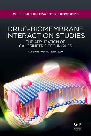
Drug-Biomembrane Interaction Studies
The Application of Calorimetric Techniques
- Rosario Pignatello(Author)
- 2013(Publication Date)
- Woodhead Publishing(Publisher)
All living cells, prokaryotic and eukaryotic, possess a thin cell or Plasma Membrane (also known as plasmalemma), which encloses their contents and acts as a semi-porous barrier to the outside environment. It also serves as the communications interface between the cell and its environment. Biological membranes also compartmentalize cellular organelles and their functions. Inside a cell, the endoplasmic reticulum, Golgi apparatus, lysosomes, vesicles and vacuoles are surrounded by a single biomembrane sheet. Mitochondria and the nucleus are surrounded by two membrane layers. Finally, the membrane regulates the flow of materials into and out of the cell, mediates intercellular communication, contacts and adhesion, and performs a multitude of other tasks.Various scientific hypotheses have been used to describe the structure of Plasma Membranes and Eichman wrote a good historical description.9 Of the various models, the most accepted theory is the fluid mosaic model, which was developed by Singer and Nicolson in 1972. According to this theory, a cell membrane consists of a continuous, fluid, double layer of phospholipid (PL), containing or attaching to in different ways other components like proteins, carbohydrates and cholesterol (CHOL) (see below). In a very simplified visualization, the PLs form a thin, flexible sheet, while the proteins float in this sheet like icebergs, and the carbohydrates protrude out from the surface (Figure 1.1 ).Figure 1.1 The fluid mosaic model of cell membranesThe name of this model arises from the assumption that the Plasma Membrane is not rigid, but exists in a fluid state where the different molecules are arranged like a mosaic pattern. The arrangement of these molecules is, however, not random, but regulated (in the physiological state) or dysregulated (in a disease state), which affects the coexistence, movement, trafficking and function of each component and of the entire membrane. Alterations to cell membrane dynamics and strength are often associated with disease. eBook - PDF
Infrastructure and Activities of Cells
Biotechnology by Open Learning
- M.C.E. van Dam-Mieras, B C Currell, R C E Dam-Mieras(Authors)
- 2016(Publication Date)
- Butterworth-Heinemann(Publisher)
60 Chapter 3 The structure and function of membranes 3.1 Introduction Cells in multicellular organisms are maintained as distinct entities by the Plasma Membrane. This acts both as a barrier, separating the cell's internal solutions from those around it, and as a transport system, allowing the passage of certain compounds into or out of the cell. The functioning of this membrane is of paramount importance to the organism as a whole because the specialised functioning of cells depends upon the proper regulation of what goes in and what goes out. If cells are treated with chemicals which alter the permeability properties of the Plasma Membrane they can no longer maintain the correct internal environment and cease to function properly. In this chapter we will explore the structure of the Plasma Membrane, and indirectly, that of other cellular membranes, to see if we can understand how it carries out its various functions. 3.2 Early studies of the membrane needed a readily available source of cells If a scientist is planning to study Plasma Membranes a suitable source of material is necessary. One particular source has been used much more than any other. n Can you suggest what source of Plasma Membranes have been used more than any other? Would it be of plant or animal origin? Ideally this source will be readily available and it must be reasonably easy to obtain Plasma Membrane material from it. If the Plasma Membrane can be obtained in relatively pure form so much the better. I I Do you know of a cell type which satisfies these criteria? The answer is the human red blood cell. This is a quite unusual cell type because when it is fully mature it no longer contains a nucleus and has virtually no other internal membranes. It is also available from blood banks in considerable quantity. Thus red blood cells are a potentially useful source of membranes. Having got the cells, we now break them open by lysis, using the process of osmosis. eBook - ePub
Medicinal Chemistry
An Introduction
- Gareth Thomas(Author)
- 2011(Publication Date)
- Wiley(Publisher)
7 Biological membranes 7.1 IntroductionAll cells have a membrane, known as the cytoplasmic or Plasma Membrane , that separates the internal medium of a cell (intracellular fluid ) from its surrounding medium (extracellular fluid ). In the cells (eukaryotes ) of higher organisms, membranes also form the boundaries of the internal regions that retain the intracellular fluids in separate compartments (Figs. 7.1a and 7.1b ). Those compartments that can be recognised as separate entities, such as the nucleus, mitochondria and lysosomes, are known collectively as organelles . Organelles carry out specialised tasks within the cell. However, in the prokaryotic cells (Fig. 7.1c ) of simpler organisms where there are no organelles the Plasma Membrane is also involved in many of the functions of the organelles. The more fragile Plasma Membranes of plant cells and bacteria are also protected by a more rigid exterior covering known as the cell wall.The primary function of the Plasma Membrane is to maintain the integrity of the cell in its environment. It is now also known that the membranes of all types of cell regulate the transfer of substances in and out of the cell and between its internal compartments. This movement controls the health, as well as the flow of information between and within cells. The Plasma Membrane of a cell is also involved in both the generation and receipt of chemical and electrical signals, cell adhesion, which is responsible for tissue formation, cell locomotion, biochemical reactions and cell reproduction. The internal cell membranes have similar functions and, in addition, are often actively involved in the function of organelles. Most drugs either interact with the receptors and enzymes attached to the membrane or have to pass through a membrane in order to reach their site of action.The role of membranes and cell walls in maintaining cell integrity and their involvement in cellular function makes these areas of cells potential targets for drug action. However, in
- Wes Stein(Author)
- 2012(Publication Date)
- Academic Press(Publisher)
C H A P T E R 1 The Anatomy of the Plasma Membrane 1.1 Some Considerations of Methodology This book is concerned with the problem of how molecules and ions move across cell membranes. T o handle this problem effectively, we will need to understand in some detail the structure of cell membranes, what molecules the membrane is composed of, and how these molecules are arranged. In the present chapter we shall consider these problems but shall find that they have no easy solution. One reason for this is that, as Ponder ( 1 9 6 1 ) has pointed out, the very definition of the term cell membrane is a matter of contention. In fact, we cell biologists use the term cell membrane or plasma m e m b r a n e — w e shall use these terms interchangeably—in at least three quite different senses. In the anatomical sense, the cell membrane is the external limiting region of the cell, visible occasionally as a darkly staining region in the light microscope and with more certainty in the electron microscope as a layer (or pair of layers) of osmiophilic material. In the biochemical sense, the cell membrane is a fraction of the cell prepared b y the now classical techniques of selective disintegration of the whole cell, followed by differential centrifugation. A preparation is obtained which can b e analyzed chemically and which can, by electron microscopy, be compared with the cell membrane seen in the whole cell. Finally, in the physiological sense, the cell membrane is a hypothetical struc-ture invented to explain certain data on the permeability of cells (that is, on the rate of entry into these cells of certain substances) and to explain other data on the distribution of metabolites and other mol-ecules between the cell and the fluid in which the cell is immersed. Such data often suggest the presence of a permeability barrier be-tween the cell and its environment. eBook - PDF
- Mary Ann Clark, Jung Choi, Matthew Douglas(Authors)
- 2018(Publication Date)
- Openstax(Publisher)
5 | STRUCTURE AND FUNCTION OF Plasma MembraneS Figure 5.1 Despite its seeming hustle and bustle, Grand Central Station functions with a high level of organization: People and objects move from one location to another, they cross or are contained within certain boundaries, and they provide a constant flow as part of larger activity. Analogously, a Plasma Membrane’s functions involve movement within the cell and across boundaries' activities. (credit: modification of work by Randy Le’Moine) Chapter Outline 5.1: Components and Structure 5.2: Passive Transport 5.3: Active Transport 5.4: Bulk Transport Introduction The Plasma Membrane, the cell membrane, has many functions, but the most basic one is to define the cell's borders and keep the cell functional. The Plasma Membrane is selectively permeable. This means that the membrane allows some materials to freely enter or leave the cell, while other materials cannot move freely, but require a specialized structure, and occasionally, even energy investment for crossing. Chapter 5 | Structure and Function of Plasma Membranes 143 5.1 | Components and Structure By the end of this section, you will be able to do the following: • Understand the cell membrane fluid mosaic model • Describe phospholipid, protein, and carbohydrate functions in membranes • Discuss membrane fluidity A cell’s Plasma Membrane defines the cell, outlines its borders, and determines the nature of its interaction with its environment (see Table 5.1 for a summary). Cells exclude some substances, take in others, and excrete still others, all in controlled quantities. The Plasma Membrane must be very flexible to allow certain cells, such as red and white blood cells, to change shape as they pass through narrow capillaries. These are the more obvious Plasma Membrane functions. eBook - PDF
- Silvia Muro(Author)
- 2016(Publication Date)
- Jenny Stanford Publishing(Publisher)
Non-constitutive exocytosis and subsequent endocytosis are highly energy-expending processes and are thus dependent on mitochon-dria [60, 61]. 2.7 Conclusions and Perspectives In conclusion, the Plasma Membrane allows the delimitation of environments (intracellular space), the key to the identity of a single cell. It is also the interface for communication between cells, which carries out this task with great precision and without a lot of noise. Many drugs need to pass through one or more cell membranes to reach their site of action. Chemical and electrical properties of a Plasma Membrane can act as obstacles or gates. Investigators, membrane engineers, and factories need to evaluate these properties when building ideas, methods, and molecules for the health of living organisms. References 1. Nageli, K. W. (1887). The Microscope in Theory and Practice (Swan Sonnenschein, Lowrey, London, UK). 2. Heilbrunn, L. V. (1956). The Dynamics of Living Protoplasm (Academic Press, New York, USA). 60 Plasma Membrane as a Semipermeable Barrier 3. Davson, H., and Danielli, J. F. (1943). The Permeability of Natural Membranes (Hafner Publishing Company, New York, USA). 4. Singer, S. J., and Nicolson, G. L. (1972). The fluid mosaic model of the structure of cell membrane, Science , 175 , pp. 720–731. 5. Yeagle, P. L. (2005). The Structure of Biological Membranes , 2nd Ed. (Boca Raton, CRC Press, FL, USA). 6. Sonnino, S., and Prinetti, A. (2013). Membrane domains and the “lipid raft” concept, Curr. Med. Chem. , 20 , pp. 4–21. 7. Vaz, W. L. (1994). Diffusion and chemical reactions in phase-separated membranes, Biophys. Chem ., 50 , pp. 139–145. 8. Sheikh, K. H., and Jarvis, S. P. (2011). Crystalline hydration structure at the membrane–fluid interface of model lipid raft indicates a highly reactive boundary region, J. Am. Chem. Soc. , 133 , pp. 18296–18303. 9. Cossis, A. R., and Prosser, C. L. (1978). Evolutionary adaptation of membranes to temperature, Proc. eBook - PDF
The Plant Cell
A Comprehensive Treatise
- N. E. Tolbert(Author)
- 2013(Publication Date)
- Academic Press(Publisher)
The Plasma Membrane ROBERT T. LEONARD THOMAS K. HODGES 4 I. Introduction 163 II. Morphology as Distinguished with the Electron Microscope . 164 A. Transmission Electron Microscopy 164 B. Freeze-Fracture Electron Microscopy 169 III. Purification of the Plasma Membrane 169 A. Generalized Procedure 169 B. Identification of Plasma Membrane Vesicles in Subcellular Fractions 171 C. Contaminants in the Plasma Membrane Fraction 173 IV. Chemical Composition of the Plasma Membrane 174 A. Lipids 174 B. Proteins 174 C. Carbohydrates 175 V. Enzymatic Composition of the Plasma Membrane 175 A. K+-ATPase 175 B. Glycosyl Transferase 176 C. CeUulase 177 D. Other Functional Proteins 177 VI. Functions 178 A. Transport 178 B. Synthesis and Assembly of Cell Wall Microfibrils . . . . 180 C. Receptor for Hormonal and Environmental Signals . . . . 180 VII. Conclusions 181 References 181 1. INTRODUCTION The protoplast of plant cells, as with cells of other organisms, is separated from its environment by the Plasma Membrane or plasmalemma. The term The Biochemistry of Plants, Vol. 1 Copyright © 1980 by Academic Press, Inc. All rights of reproduction in any form reserved. ISBN 0-12-675401-2 163 164 Robert Τ. Leonard and Thomas K. Hodges II. MORPHOLOGY AS DISTINGUISHED WITH THE ELECTRON MICROSCOPE A. Transmission Electron Microscopy The Plasma Membrane, as revealed by conventional transmission electron microscopy (Fig. 1), appears as a thin undulating Une around the periphery plasmalemma (''lemma, the shell of a fruit) is often used to refer to the surface membrane of the plant cell, but here we will refer to this membrane as the Plasma Membrane. Early attempts to verify the existence of the Plasma Membrane in plants were confounded by the presence of the thick and resistant cell wall. It was necessary to separate the Plasma Membrane from the cell wall by plasmolysis to conduct experiments designed to demon-strate the presence of an outer cell membrane. eBook - ePub
- Subrata Pal(Author)
- 2019(Publication Date)
- Academic Press(Publisher)
Chapter 14Membrane structure and function
Abstract
Biomembranes consist of a hydrophobic matrix formed by a lipid bilayer with surface-bound and embedded proteins. The bilayer is both diverse and asymmetric in respect to its lipid composition. However, the amphipathic nature of all kinds of lipid molecules present in the membrane thermodynamically favors the formation of the common bilayer structure. Both lipids and proteins in the bilayer are in constant motion. As the boundary of a cell, the membrane displays a property of selective permeability, allowing only certain substances to be passively or actively transported across it. It also plays a crucial role in the cell’s response to environmental signals.Keywords
Biomembrane; Lipid bilayer; Membrane protein; Fluid mosaic model; Transport across membrane; Signal transductionIn order to sustain life, cells need to carry out a wide variety of chemical reactions. Earlier, we have discussed the synthesis of some of the most important biomolecules—DNA, RNA, and protein. However, we have to understand that the cell is able to synthesize its molecular constituents as well as carry out a number of other metabolic processes, efficiently and in an organized manner, since it is partially secluded from its more random environment by a biomembrane. The hydrophobic core of the membrane is formed by lipid assemblies.14.1 Composition of the membrane
In a living cell, lipids perform three general functions: (a) storage of energy as triglycerol ester and steryl esters, (b) as first and second messengers in signal transduction and molecular recognition processes, and (c) formation of the matrix of cellular membranes. It is the third function that we are concerned with in this chapter.Some bacterial cells (Gram-positive) contain just one membrane, whereas others (Gram-negative) are surrounded by two membranes—inner and outer. Eukaryotic cells are enclosed in a single Plasma Membrane. Some of the internal organelles of eukaryotic cells such as the nucleus, mitochondria, and chloroplasts are surrounded by double lipid bilayers, while endoplasmic reticula, Golgi apparatus, and lysosomes each contains a single lipid bilayer. Nevertheless, in all cases, lipids form the common core. Besides, the cell membranes also contain proteins and carbohydrates of different types and in varying amounts. eBook - ePub
- Andrey B. Rubin(Author)
- 2017(Publication Date)
- Wiley-Scrivener(Publisher)
Chapter 12 Molecular Organization of Biological Membranes12.1 Composition and Structure of Biological Membranes
Biological membranes are functional cell structures with the thickness of several molecular layers enclosing the cytoplasm and most of intracellular structures and forming a uniform intracellular system of small channels, folds and closed cavities. The thickness of biological membranes is occasionally more than 10.0 nm, but due to a relatively tight packing of their basic molecular components (proteins and lipids), as well as large overall area of cell membranes as a rule, they make more than half of the dry cell mass.Biological membranes consist mostly of proteins, lipids and carbohydrates.Proteins and lipids make the most part of the mass of dry membranes. Usually, the portion of carbohydrates does not exceed 10–15 %, they being linked either to protein molecules (glycoproteins) or to lipid molecules (glycolipids). In membranes of different origin, the content of lipids varies from 25 to 75 % relative to protein mass (Table 12.1 ).Table 12.1. Content of Lipids in Mammalian Cell Membranes, % of the Mass of All LipidsLipids Plasma Membranes Mitochondrii Lysosomes Nuclei Endoplasmic reticulum Golgi apparatus Phosphatidylcholine 18.5 37.5 23.0 44.0 48.0 24.5 Sphingomyelin 12.0 0 23.0 3.0 5.0 6.5 Phosphatidylethanolamine 11.5 28.5 12.5 16.5 19.0 9.0 Phosphatidylserine 7.0 0 6.0 3.5 4.0 2.5 Phosphatidylinositol 3.0 2.5 6.0 6.0 7.5 5.0 Lysophosphatidylcholine 2.5 0 0 1.0 1.5 3.0 Diphosphatidyl glycerol  eBook - ePub
Netter's Essential Physiology E-Book
Netter's Essential Physiology E-Book
- Susan Mulroney, Adam Myers(Authors)
- 2015(Publication Date)
- Elsevier(Publisher)
Fig. 1.2 ).Figure 1.2 Buffering the External EnvironmentIn multicellular organisms, the basic homeostatic mechanisms of single-celled organisms are mirrored by integration of specialized organ systems to create a stable environment for the cells. This system allows specialization of cellular functions and a layer of protection for the systems.The Cell Membrane
The human body is composed of eukaryotic cells (those that have a true nucleus) containing various organelles (e.g., mitochondria, smooth and rough endoplasmic reticulum, Golgi apparatus) that perform specific functions. The cell, with its nucleus and organelles, is surrounded by a Plasma Membrane consisting of a lipid bilayer primarily made of phospholipids, with varying amounts of glycolipids, cholesterol, and proteins. The lipid bilayer is positioned with the hydrophobic fatty acid tails of phospholipids oriented toward the middle of the membrane and the hydrophilic polar head groups oriented toward the extracellular or intracellular space. The fluidity of the membrane is maintained in large part by the amount of short-chain and unsaturated fatty acids incorporated within the phospholipids; incorporation of cholesterol into the lipid bilayer reduces fluidity (Fig. 1.3 ). The oily, hydrophobic interior region makes the bilayer an effective barrier to fluid (on either side), with permeability only to some small hydrophobic solutes, such as ethanol, that can diffuse through the lipids.Figure 1.3 The Eukaryotic Plasma MembraneThe Plasma Membrane is a lipid bilayer, with hydrophobic ends oriented inward and hydrophilic ends oriented outward. Primary constituents of the membrane are phospholipids, glycolipids, and cholesterol. A wide variety of proteins are associated with the membrane, including (1) ion channels, (2) surface antigens, (3) receptors, and (4) eBook - PDF
- Gerald Karp, Janet Iwasa, Wallace Marshall(Authors)
- 2018(Publication Date)
- Wiley(Publisher)
(b) The same experiment shown in a is carried out for 33 ms in an artificial bilayer, which lacks the “picket fences” present in a cellular membrane. The much more open, extended trajectory of the phospholipid can now be explained by simple, unconfined Brownian movement. For the sake of comparison, fake compartments were assigned in b and indicated by different colors. SOURCE: From Takahiro Fujiwara et al., Courtesy of Akihiro Kusumi, Nagoya, J. Cell Biol. 157:1073, 2002; Reproduced with permission of The Rockefeller University Press. 8.7 • The Dynamic Nature of the Plasma Membrane 333 334 CHAPTER 8 • Cellular Membrane 8.8 The Red Blood Cell: An Example of Plasma Membrane Structure Of all the diverse types of membranes, the Plasma Membrane of the human erythrocyte (red blood cell) is the most studied and best understood (FIGURE 8.32a). There are several reasons for the popularity of this membrane. The cells are inexpensive to obtain and readily available in huge numbers from whole blood. They are already present as single cells and need not be dissoci- ated from a complex tissue. The cells are simple by comparison with other cell types, lacking nuclear and cytoplasmic mem- branes that inevitably contaminate Plasma Membrane prepara- tions from other cells. In addition, purified, intact erythrocyte Plasma Membranes can be obtained simply by placing the cells in a dilute (hypotonic) salt solution. The cells respond to this osmotic shock by taking up water and swelling, a phenomenon termed hemolysis. As the surface area of each cell increases, the cell becomes leaky, and the contents, composed almost totally of dissolved hemoglobin, flow out of the cell leaving behind a Plasma Membrane “ghost” (Figure 8.32b). Once erythrocyte Plasma Membranes are isolated, the pro- teins can be solubilized and separated from one another (frac- tionated), providing a better idea of the diversity of proteins within the membrane. eBook - PDF
Karp's Cell and Molecular Biology
Concepts and Experiments
- Gerald Karp, Janet Iwasa, Wallace Marshall(Authors)
- 2016(Publication Date)
- Wiley(Publisher)
Treatment of the membrane with agents that disrupt the underlying membrane skeleton removes some of the fences that restrict phospholipid diffu- sion. But if the membrane skeleton lies beneath the lipid bilayer, how can it interfere with phospholipid movement? The authors of these studies speculate that the fences are constructed of rows of integral membrane proteins whose cytoplasmic domains are attached to the membrane skeleton. This is not unlike the confinement of horses or cows by a picket fence whose posts are embedded in the underlying soil. MEMBRANE DOMAINS AND CELL POLARITY For the most part, studies of membrane dynamics, such as those discussed above, are carried out on the relatively homogeneous plasma mem- brane situated at the upper or lower surface of a cell residing on a cul- ture dish. Most membranes, however, exhibit distinct variations in protein composition and mobility, especially in cells whose various surfaces display distinct functions. For example, the epithelial cells that line the intestinal wall or make up the microscopic tubules of the kid- ney are highly polarized cells whose different surfaces carry out differ- ent functions (FIGURE 4.30). Studies indicate that the apical Plasma Membrane, which selectively absorbs substances from the lumen, pos- sesses different enzymes than the lateral Plasma Membrane, which interacts with neighboring epithelial cells, or the basal membrane, which adheres to an underlying extracellular substrate (a basement membrane). In other examples, the receptors for neurotransmitter substances are concentrated into regions of the Plasma Membrane located within synapses (see Figure 4.57), and receptors for low‐density lipoproteins are concentrated into patches of the Plasma Membrane specialized to facilitate their internalization (see Figure 8.38). Of all the various types of mammalian cells, sperm may have the most highly differentiated structure.
Index pages curate the most relevant extracts from our library of academic textbooks. They’ve been created using an in-house natural language model (NLM), each adding context and meaning to key research topics.