Biological Sciences
Slime Mold
Slime molds are a group of organisms that display characteristics of both fungi and protozoa. They are single-celled organisms that can aggregate to form multicellular structures. Slime molds are known for their unique life cycle, which includes a mobile feeding stage and a stationary reproductive stage. They play important roles in decomposition and nutrient cycling in ecosystems.
Written by Perlego with AI-assistance
Related key terms
1 of 5
8 Key excerpts on "Slime Mold"
 eBook - PDF
- John Webster, Roland Weber(Authors)
- 2007(Publication Date)
- Cambridge University Press(Publisher)
2 Protozoa: Myxomycota (slime moulds) 2.1 Introduction When the first slime moulds were described by Johann H. F. Link in 1833, they were given the term myxomycetes (Gr. myxa ¼ slime). Link used the suffix -mycetes because of the superficial similarity of the fructifications of slime moulds with the fruit bodies of certain fungi, notably Gasteromycetes (see Chapter 20). Although it has been appreciated for some time that they lack any true relationship with the Eumycota (de Bary, 1887; Whittaker, 1969), slime moulds have none the less been studied mainly by mycologists rather than protozoologists, prob-ably because they occur in the same habitats as fungi and are routinely encountered during fungus forays. Since slime moulds are only rarely covered by zoology courses even today, they are briefly described in this chapter, referring to more specialized literature as appropriate. Slime moulds differ substantially from the Eumycota not only in phylogenetic terms, but also regarding their physiology and eco-logy. Their vegetative state is that of individual amoebae in the cellular slime moulds, or of a multinuclear (coenocytic) plasmodium in the plasmodial slime moulds. Motile stages bearing usually two anterior whiplash-type flagella may be present in the plasmodial slime moulds (Sections 2.4, 2.5) and in the Plasmodiophoromy-cota (Chapter 3). Amoebae or plasmodia feed by the ingestion ( phagocytosis ) of bacteria, yeast cells or other amoebae. This is followed by intracellular digestion in vacuoles. The mode of nutrition in slime moulds is therefore fundamen-tally different from extracellular degradation and absorption as shown by Eumycota. Numerous phylogenetic analyses of DNA sequences encoding rRNA molecules and various structural proteins or enzymes have been carried out, but the results obtained are difficult to interpret because the comparison of different genes have led to rather variable phylogenetic schemes.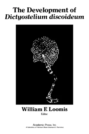 eBook - PDF
- William Loomis(Author)
- 2012(Publication Date)
- Academic Press(Publisher)
This was shown in a well-known 1940 paper by Raper, which was followed by many additional papers from his laboratory over the years. What I have called the modern era has a number of trends, all of which are reflected in this volume. Some researchers have been concerned with the discovery of new species, with ecological problems, and with problems in-volving the relationship of different major groups of Slime Molds to one another. Others have been concerned with the traditional problems of de-velopmental biology and have experimented on the cellular level. Still oth-ers have concentrated on various important aspects of the biochemistry of Slime Mold development. This molecular approach was given a great boost by the development of axenic strains, thereby eliminating confusion of the chemistry of the bacterial food supply (R. Sussman and M. Sussman, 1967; Ashworth and Watts, 1970; Loomis, 1971). In most studies there has been a combination of these two methods, and they have included areas such as genetics, gene action, Chemotaxis, cell adhesion, and differenct aspects of 1 . Comparative Biology of Cellular Slime Molds 3 differentiation including pattern formation. There has also been consider-able interest in the physiology of phototropism and thermotropism. The reason for such an enormous recent profusion of studies on all these different aspects of cellular Slime Mold development arises from the conviction that these organisms are ideal for the study of fundamental problems of develop-ment. They are multicellular and eukaryotic and have a simple mor-phogenesis. To some extent we have all been consciously or unconsciously influenced by the success of Ε. coli in molecular biology and hope that the cellular Slime Molds will illuminate the next level of complexity. II. The Life History As has been done so often, I will illustrate the unusual feature of the development by outlining the life history of Dictyostelium discoideum .
- Gabrielle I. Edwards, Cynthia Pfirrmann, Barron's Educational Series(Authors)
- 2021(Publication Date)
- Barrons Educational Services(Publisher)
Many other protist species are helpful to humans. Slime Molds help to keep an ecological balance by feeding on decayed plant and animal matter. These organisms also have a great deal of value as research specimens for investigators who are trying to ferret out the secrets of cell specialization and differentiation.Plankton is the mass of green that floats on rivers, lakes, and oceans. In reality, protists—microscopic “plant” life (phytoplankton) and microscopic “animal” life (zooplankton)—intermingle with mutual benefit in this floating mass, which serves as vital roles in the food chains of aquatic species.Review Exercises
WORD-STUDY CONNECTION
chytrid cilia colony conjugation contractile vacuole cyst diatom diatomaceous earth dinoflagellate fungus hypochytrid macronucleus micronucleus nematocyst oral groove paramylum pellicle plankton plasmodium protest protozoa pseudoplasmodium pseudopod pyrenoid body rhizoid Slime Mold social amoeba sorocarp sporangium stigma symbiosis trichocysts tsetse zooflagellate zooplankton zoospore zygoteSELF-TEST CONNECTIONPART A. Completion. Write in the word that correctly completes the statement.1 .One-celled protists that resemble animal cells are the _____.2 .Another name for a “self-feeder” is a (an) _____.3 .Amoebae move by means of false feet known as _____.4 .The usual mode of reproduction in protozoa is _____.5 .The relationship in which two organisms of different species live together and neither is harmed by the association is known as _____.6 .Digestion in Amoeba proteus takes place in the _____.7 .The Sporozoa are harmful to organisms of other species and are therefore classified as _____.8 .Anopheles is the genus name of a _____.9 .The organelle that expels excess water from the protist is the _____.10 .In Paramecium, the nucleus that controls metabolic activity is the _____.11 .The body of the paramecium is prevented from being totally flexible by the _____.12 eBook - ePub
Thorp and Covich's Freshwater Invertebrates
Ecology and General Biology
- James H. Thorp, D. Christopher Rogers(Authors)
- 2014(Publication Date)
- Academic Press(Publisher)
Cavostelium ). The dictyostelids are typically phagotrophic amebae; but when starved, they aggregate, form a migrating slug, and then produce a fruiting body from which spores are released. These later germinate to produce amebae. In the myxomycetes, individual amebae (sometimes thousands of them) with or without flagella coalesce to form a distinctive (often brightly colored) multinucleate plasmodium that gives rise to fruiting bodies. Slime Molds are common in damp forest soils, tree bark, and dead or dying wood, and abundance and local species richness are greater in deciduous than in coniferous forests. There is extreme patchiness in the distribution of dictyostelid species in forest soils, and coexistence of multiple species in the same small soil samples is rare. Most myxomycete species are believed to be cosmopolitan. The phagotrophic ameboid stages of Slime Molds may be quantitatively important grazers of bacteria, fungi, and the other primary decomposers of organic matter in soil. Other ecological interactions of Slime Molds may be at least as complex as their life cycles (e.g., the migrating slugs of dictyostelids appear capable of repelling grazing nematodes).Flagellated Protozoa
There is little consensus on how to classify the flagellates, and they are here divided into broad functional groups (Figures 7.1 and 7.3 ). Heterotrophic, nonphotosynthetic flagellates are fundamentally important because they are abundant (there are seldom fewer than 100/ml, sometimes 1000/ml, even in the plankton) and because their grazing activities are largely responsible for controlling the abundance of bacteria in aquatic environments. In some taxonomic groups, all species are exclusively heterotrophic (e.g., choanoflagellates and bodonids); others contain many mixotrophs (e.g., the euglenids and chrysomonads), whereas the haptomonads and cryptomonads are dominated by phototrophs and only a minority are capable of phagotrophy. In the past 25 years, a large diversity of heterotrophic flagellates has been discovered. Most of these are choanoflagellates, chrysomonads, euglenids, or bodonids. Some of the more easily recognized species (e.g., Rhynchomonas nasuta ) have been recorded from a wide range of habitat types in freshwater, marine, and terrestrial environments (Patterson and Larsen, 1991 ; Lee and Patterson, 1998 eBook - PDF
Networks Of Interacting Machines: Production Organization In Complex Industrial Systems And Biological Cells
Production Organization in Complex Industrial Systems and Biological Cells
- Dieter Armbruster, Kunihiko Kaneko, Alexander S Mikhailov(Authors)
- 2005(Publication Date)
- World Scientific(Publisher)
The transduction of sensed information, integration of multiple signals, decision making, biocomputing, emergence of the rhythm, order among multiple rhythms, allometry in locomotion, etc. are described from a viewpoint of transitions among dynamic dissipative structures on a metabolic network 221 222 T. Ueda or in a collective dynamics of coupled oscillators. 112,334,5 9.1.1. The T h e Slime Molds, Like Nothing on Earth The true Slime Molds are eukaryotic micro-organisms. They are unique not because they have solitary features in taxonomic classification, but because they share common features with all the other four kingdoms of fungus, protozoan, plant and animal. So the taxonomists more than a century ago named the organisms appropriately myxomycetes (slime + fungus), or myc- etazoa (fungus + animal). Phytochromes, known as photosensors in higher plants, are also found in this organism, suggesting a link to plants. The organism reacts behaviorally, performing high degree of information pro- cessing like that of brain in higher animals. These latter two are our main topics in this chapter. The true Slime Molds are cosmopolitan and free-living fed on rotten logs and fallen leaves in forests all around the world. The rich variety in the morphology of sporangia and spores is a basis of classifica- tion of this group, and the number of species discovered is now reaching one thousand All share the common life cycle, as shown in Fig. 9.1. The myxoamoeba germinates from spores, and proliferates as a single nuclear cell by feeding with bacteria. Although all the myxoamoebae look similar, there is a sex differentiation. Two haploid amoebae with different sex types conjugate to form a diploid zygote. This is the beginning of the unusual life of plasmodium. Here unlike other living organisms, the nuclear division, spomngeum j& genriimtion plasmodium z Fig. 9.1. The life cycle of the true Slime Mold Physarum polycephalum. eBook - PDF
Life at the Edge of Sight
A Photographic Exploration of the Microbial World
- Scott Chimileski, Roberto Kolter(Authors)
- 2017(Publication Date)
- Belknap Press(Publisher)
115 4 Intelligent Slime IT’S NIGHTTIME ON THE TOP FLOOR of a life sciences research center in the city of Boston. There are no voices or footsteps in the hallways, only the machine noise of ventilation fans, shaking cultures, and whistling steam pipes. Hundreds of mi-crobial species grow, cell by cell, inside petri plates stacked in dark incubators. But one microbe in the building is behaving differently. Nearby, a species name is written in fresh ink: Physarum polycephalum, the many-headed Slime Mold. Inside of the petri plate, a network of bright yellow tubes is moving across the semi-solid agar medium. Each tube pulsates like a vein, expanding to twice its width and contracting to its original size once every minute. The tip of each tube extends into fan-shaped sheets that act like feet. Pulling itself forward, it creeps along centimeter by centimeter. Behind the extensions at the leading edge, tubes connect to form a gooey web. It appears to be a life-form out of a science-fiction story, an organism tele-ported from a planet in a distant galaxy. It is, nevertheless, an amoeba: a eukary-otic microbe that evolved right here on Earth over a billion years ago, colonizing land before any plant or animal. However, Physarum is not like other microbes. Most of the time, it’s not a microbe at all. One day, it might be far too small to see, hidden within leaf litter on the forest floor. The next day, if it encounters enough bacteria or fungi, it becomes a voracious predator, rapidly growing into a mac-roscopic slime called a plasmodium that can cover an area larger than a square meter. It transitions from invisible flagellated amoeboid cells through the visible feeding and fruit body stages, then back to invisible reproductive spores that can lay dormant and then germinate decades later. Its life cycle is situated directly above and below the limits of the human eye, traversing the micro and macro realms. eBook - PDF
Cell Biology of Physarum and Didymium V2
Differentiation, Metabolism, and Methodology
- Henery Aldrich(Author)
- 2012(Publication Date)
- Academic Press(Publisher)
PART I Differentiation This page intentionally left blank CHAPTER 1 Developmental Biology of Slime Molds—An Overview ROLAND J. WICK and HELMUT W. SAUER I. Introduction 3 II. Life as a Plasmodium 4 A. Formation of Macrocysts 8 B. Sporulation 11 III. Amoeba-Flagellates 14 A. Formation of Microcysts 15 B. Amoeba-Plasmodium Transition 15 References 17 I . I N T R O D U C T I O N Myxomycetes are truly remarkable organisms in that they are more than uni-cellular yet less than multicellular but are clearly eukaryotic systems. Therefore, the analysis of their life cycles, from single cells, microscopic myxamoebae, or flagellates, to macroscopic multinucleated plasmodia and fruiting bodies, and back to the unicellular spores, has become a target for researchers from almost every discipline of the biosciences. Although myxomycetes deserve to be studied in their own right, in most of the recent biochemical and experimental work, they are considered 4 'model sys-tems, from which an understanding of the two basic problems in cell biology—growth and differentiation—might be reached. In this chapter, we describe some aspects of developmental biology, which have been previously reviewed by von Stosch (1965), Alexopoulos (1966), Sauer (1973), Collins (1979), and Sachsenmaier (1979), and most extensively by Gorman and Wilkins (1980). Nevertheless, despite the large body of information available, the main devel-opmental reactions are still not resolved; further investigations, which would 3 CELL BIOLOGY OF PHYSARUM AND DIDYMIUM, VOL. II Copyright © 1982 by Academic Press, Inc. All rights of reproduction in any form reserved. ISBN 0-12-049602-X 4 Roland J. Wick and Helmut W. Sauer utilize genetic and biochemical methods in meaningful experiments at the appro-priate stages of the life cycle, are needed. eBook - PDF
- Kenneth Bryan Raper(Author)
- 2014(Publication Date)
- Princeton University Press(Publisher)
However, as if compensating for this de- ficiency, many of these structures arise from a single primary aggregate and some perforce find the proper balance of substrate, temperature, and humidity required for sorocarp formation. The additional characteristic of producing not one but several adherent sorocarps with divergent son en- hances spore dispersal. This tendency is even further developed in species of Polysphondylium, with their whorls of sorus-tipped branches that project outward from the main stems, which are themselves constructed toward the light and into the more open areas peripheral to their sites of origin. And finally, in Acytostelium a pattern has emerged wherein no cells are sacrificed in sterile supportive tissue but all myxamoebae are transformed into reproductive spores. Sensitive to their environment and capable of singular collaboration and differentiation at the cellular level, these Slime Molds seem to have mastered the problems of when to produce their spores and how to ensure maximal distribution. There are in addition two other modes of survival, microcysts and mac- rocysts. The first represent unaggregated myxamoebae that encyst indi- vidually, hence are able to carry the Slime Molds through limited periods 414-EPILOGUE of adversity. The second are something special! They are formed under conditions often suboptimal for sorocarp formation and, insofar as we know, are without parallel in other organisms, since they arise from newly formed zygotes that first attract and then engulf neighboring myxamoebae and in the process lay down three protective walls, one of which is rich in cellulose. Unique in origin and structure, they are also the sites of sexual reproduction in the dictyostelids. The cellular Slime Molds are simple organisms by any standard one may wish to apply, and the information to be gained from them may likewise have limited application in terms of more complex and highly organized creatures.
Index pages curate the most relevant extracts from our library of academic textbooks. They’ve been created using an in-house natural language model (NLM), each adding context and meaning to key research topics.