This fully revised new edition of the classic reference on domestic animal physiology provides detailed descriptions of animal function and dysfunction, with an emphasis on clinical relevance and pedagogical features to enhance learning.
• Presents in-depth, comprehensive descriptions of domestic animal function and dysfunction • Emphasizes clinical relevance, with clinical correlations, notes of relevance, and self-assessment questions featuring situations likely to be faced in practice • Offers pedagogical features, including chapter outlines and introductions, key terms throughout the book, additional images, questions to enhance learning, and self-assessment exercises • Distills the most useful information for ease of use, with improved continuity and reduced repetition • Includes a companion website offering review questions and answers and the figures from the book in PowerPoint
Trusted by 375,005 students
Access to over 1.5 million titles for a fair monthly price.
30 The Heart and Vasculature: Gross Structure and Basic Properties
Dean H. Riedesel and Richard L. Engen
Iowa State University, Ames, IA, USA
The heart and a vast array of blood vessels that vary in size and tissue composition form the cardiovascular system. The function of the cardiovascular system can be simplified to that of a transportation system which distributes oxygen and nutrients to the tissues and removes carbon dioxide and other metabolic byproducts. The interstitial fluid environment surrounding the cells of an animal’s body must remain relatively “constant” and maintaining this consistency is known as homeostasis. The role of the cardiovascular system in homeostasis cannot be overlooked. In addition to oxygen and nutrients, hormones, white blood cells, platelets, electrolytes, and heat are also transported by the cardiovascular system and closely controlled for maintaining homeostasis. Although the cardiovascular system does not control these variables, it is used for the transportation and distribution of essential substances and byproducts that diffuse between the dense capillary networks and the interstitial fluid of tissues.
William Harvey described the mammalian circulatory system in 1628. Since that time much work has been done to study the system and research continues to this day. Although simple in principle, the cardiovascular system must have the ability to alter organ perfusion rapidly. For example, a resting skeletal muscle does not require much blood flow but as soon as it starts to exercise the need for oxygen and glucose increases rapidly. Thus, changes in the total flow of blood (cardiac output) and its distribution within the body will have to change to meet those demands. The mechanical and physiologic factors which cause and control the flow of blood in the body are better understood when basic principles of hemodynamics are reviewed. Although the direct application of physics concerning fluid flow in rigid tubes is not appropriate, the concepts are useful in understanding blood flow.
Gross structure
Describe the location of the heart in the chest cavity.
Is the heart free to move or is it held in a fixed position?
The heart is in the thoracic cavity within the mediastinum between the left and right pleural cavities and protected by the ribs from about the third to the sixth intercostal spaces. The dorsal aspect is horizontally in line with the middle of the first rib and the ventral aspect is on the sternum. The long axis of the cardiac silhouette is oriented vertically in the horse, almost vertically in ruminants, and progressively more obliquely in the pig, dog, and cat. The dorsal part of the heart is known as the base and is formed by the atria and the major vessels entering (veins) and leaving (arteries) the heart. The major vessels tend to hold the heart in a relatively fixed position dorsally while ventrally it is free within the pericardial sac.
Cardiovascular system
Describe the arterial and venous systems as being high resistance or high capacitance and explain why.
Which organ receives more blood flow, the kidney or the myocardium?
What percent of a dog’s body weight is blood?
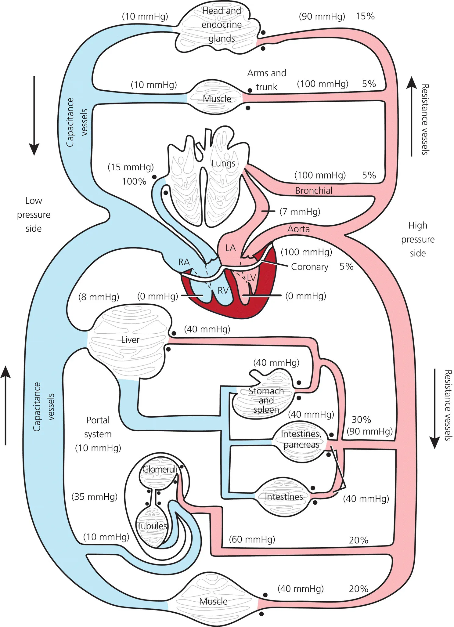
The cardiovascular system has two circulations in series: (i) the pulmonary circulation composed of the right atrium (RA), right ventricle (RV) and lungs; and (ii) the systemic circulation composed of the left atrium (LA), left ventricle (LV), and the systemic organs. Each circulation has three major divisions: (i) the distribution system (ventricles, arteries, and arterioles), (ii) the perfusion/exchange system (capillaries), and (iii) the collecting system (venules, veins, and atria). The major components of the cardiovascular system are shown in Figure 30.1, with the arterial (high-pressure) system on the right and venous (low-pressure) system on the left. The areas shaded blue represent the venous and the pulmonary arterial systems that carry blood with reduced oxygen content. The areas shaded red include the arterial and pulmonary venous systems that carry oxygenated blood. The pulmonary and systemic systems are in series such that blood flow to the lungs from the right ventricle equals blood flow in the aorta coming from the left ventricle. The amount of blood pumped by the right or left ventricle is called the cardiac output (
) and is measured in liters per minute. The distribution of blood flow is indicated (see Figure 30.1) by percentages on the arteries supplying the various organs (e.g., 15% of the cardiac output goes to the head and endocrine glands). The intravascular pressures are measured in millimeters of mercury (mmHg) and are shown (see Figure 30.1) as mean values in parentheses on both the arterial and venous systems (e.g., the mean blood pressure in the aorta is 100 mmHg).
Figure 30.1 Overview of the cardiovascular system. The blue areas represent the venous blood with reduced oxygen content; the red vessels represent the arterial system with oxygenated blood. The solid black circles indicate areas of resistance, and the percentages indicate the proportion of cardiac output delivered to the organ system at rest. ...
Table of contents
Cover
Title page
Table of Contents
List of Contributors
Preface
Acknowledgments
Tributes to Drs H. Hugh Dukes and Melvin J. Swenson
About the companion website
SECTION I: Neurophysiology
SECTION II: Body Fluids and Homeostasis
SECTION III: The Kidneys and Urinary System
SECTION IV: Respiration
SECTION V: Muscle Physiology
SECTION VI: The Cardiovascular System
SECTION VII: Digestion, Absorption, and Metabolism
SECTION VIII: Minerals, Bones, and Joints
SECTION IX: Endocrinology, Reproduction, and Lactation
Index
End User License Agreement
Frequently asked questions
Yes, you can cancel anytime from the Subscription tab in your account settings on the Perlego website. Your subscription will stay active until the end of your current billing period. Learn how to cancel your subscription
No, books cannot be downloaded as external files, such as PDFs, for use outside of Perlego. However, you can download books within the Perlego app for offline reading on mobile or tablet. Learn how to download books offline
Perlego offers two plans: Essential and Complete
Essential is ideal for learners and professionals who enjoy exploring a wide range of subjects. Access the Essential Library with 800,000+ trusted titles and best-sellers across business, personal growth, and the humanities. Includes unlimited reading time and Standard Read Aloud voice.
Complete: Perfect for advanced learners and researchers needing full, unrestricted access. Unlock 1.5M+ books across hundreds of subjects, including academic and specialized titles. The Complete Plan also includes advanced features like Premium Read Aloud and Research Assistant.
Both plans are available with monthly, semester, or annual billing cycles.
We are an online textbook subscription service, where you can get access to an entire online library for less than the price of a single book per month. With over 1.5 million books across 990+ topics, we’ve got you covered! Learn about our mission
Look out for the read-aloud symbol on your next book to see if you can listen to it. The read-aloud tool reads text aloud for you, highlighting the text as it is being read. You can pause it, speed it up and slow it down. Learn more about Read Aloud
Yes! You can use the Perlego app on both iOS and Android devices to read anytime, anywhere — even offline. Perfect for commutes or when you’re on the go. Please note we cannot support devices running on iOS 13 and Android 7 or earlier. Learn more about using the app
Yes, you can access Dukes' Physiology of Domestic Animals by William O. Reece in PDF and/or ePUB format, as well as other popular books in Medicine & Veterinary Medicine. We have over 1.5 million books available in our catalogue for you to explore.