
eBook - ePub
Obstetric Imaging: Fetal Diagnosis and Care E-Book
Joshua Copel, Mary E. D'Alton, Helen Feltovich, Eduard Gratacos, Anthony O. Odibo, Lawrence Platt, Boris Tutschek
This is a test
Compartir libro
- 848 páginas
- English
- ePUB (apto para móviles)
- Disponible en iOS y Android
eBook - ePub
Obstetric Imaging: Fetal Diagnosis and Care E-Book
Joshua Copel, Mary E. D'Alton, Helen Feltovich, Eduard Gratacos, Anthony O. Odibo, Lawrence Platt, Boris Tutschek
Detalles del libro
Vista previa del libro
Índice
Citas
Información del libro
Richly illustrated and comprehensive in scope, Obstetric Imaging, 2nd Edition, provides up-to-date, authoritative guidelines for more than 200 obstetric conditions and procedures, keeping you at the forefront of this fast-changing field. This highly regarded reference covers the extensive and ongoing advances in maternal and fetal imaging in a concise, newly streamlined format for quicker access to common and uncommon findings. Detailed, expert guidance, accompanied by superb, high-quality images, helps you make the most of new technologies and advances in obstetric imaging.
- Features more than 1, 350 high-quality images, including 400 in color.
- Helps you select the best imaging approaches and effectively interpret your findings with a highly templated, bulleted, at-a-glance organization.
- Reflects all the latest developments in the field, including genetics, open fetal surgery, fetal echocardiography, Zika virus, and 3D imaging, so you can provide the safest and most responsive care to both mother and fetus.
- Includes new chapters on Limbs and Bones Overview; Open Fetal Surgery; Biophysical Profile; Ultrasound Physics; Elastography; Doppler; MRI; Echogenic Bowel; Pregnancy of Unknown Location (PUL), Failed Pregnancy and Ectopic Pregnancy, Cesarean Scar Pregnancy; Cytomegalovirus (CMG), Rubella, Toxoplasmosis, Herpes, Varicella; and Congenital Syphilis; plus a new chapter on Zika Virus written by imaging experts from the "hot zone."
- Keeps you up to date with the latest developments in multimodality imaging and optimizing diagnostic accuracy from ultrasound, 3D ultrasound, Doppler, MRI, elastography, image-guided interventions, and much more.
Preguntas frecuentes
¿Cómo cancelo mi suscripción?
¿Cómo descargo los libros?
Por el momento, todos nuestros libros ePub adaptables a dispositivos móviles se pueden descargar a través de la aplicación. La mayor parte de nuestros PDF también se puede descargar y ya estamos trabajando para que el resto también sea descargable. Obtén más información aquí.
¿En qué se diferencian los planes de precios?
Ambos planes te permiten acceder por completo a la biblioteca y a todas las funciones de Perlego. Las únicas diferencias son el precio y el período de suscripción: con el plan anual ahorrarás en torno a un 30 % en comparación con 12 meses de un plan mensual.
¿Qué es Perlego?
Somos un servicio de suscripción de libros de texto en línea que te permite acceder a toda una biblioteca en línea por menos de lo que cuesta un libro al mes. Con más de un millón de libros sobre más de 1000 categorías, ¡tenemos todo lo que necesitas! Obtén más información aquí.
¿Perlego ofrece la función de texto a voz?
Busca el símbolo de lectura en voz alta en tu próximo libro para ver si puedes escucharlo. La herramienta de lectura en voz alta lee el texto en voz alta por ti, resaltando el texto a medida que se lee. Puedes pausarla, acelerarla y ralentizarla. Obtén más información aquí.
¿Es Obstetric Imaging: Fetal Diagnosis and Care E-Book un PDF/ePUB en línea?
Sí, puedes acceder a Obstetric Imaging: Fetal Diagnosis and Care E-Book de Joshua Copel, Mary E. D'Alton, Helen Feltovich, Eduard Gratacos, Anthony O. Odibo, Lawrence Platt, Boris Tutschek en formato PDF o ePUB, así como a otros libros populares de Medizin y Medizintechnik & Zubehör. Tenemos más de un millón de libros disponibles en nuestro catálogo para que explores.
Información
Part 1
Atlas of Selected Normal Images
1
Atlas of Selected Normal Images
Mert Ozan Bahtiyar, Carole Gravino
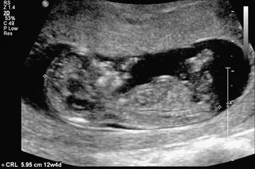
Fig. 1.1 The crown-rump length is a measurement of the length of human fetuses from the top of the crown to the bottom of the rump. It is used to estimate gestational age.
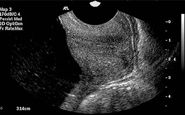
Fig. 1.2 Transvaginal ultrasound and sagittal long-axis view of the endocervical canal. Both the internal os and the external os are well visualized. The cervical length is measured from the internal os to the external os along the endocervical canal.
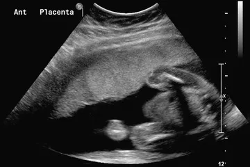
Fig. 1.3 Sagittal view of the uterus with an anterior (Ant) placenta.
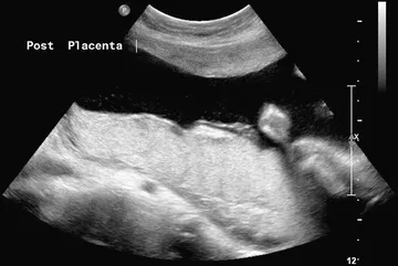
Fig. 1.4 Sagittal view of the uterus showing a posterior (Post) placenta.
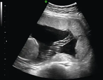
Fig. 1.5 Normal umbilical cord insertion into the placenta.
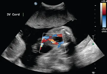
Fig. 1.6 Fetal umbilical cord insertion site. The umbilical arteries emerge caudally—originating at the iliac arteries and coursing along the margin of the urinary bladder. The umbilical vein proceeds cephalad and joins the fetal portal circulation. 3V, Three-vessel.
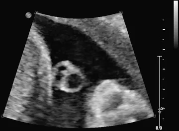
Fig. 1.7 Transverse view of the umbilical cord. The umbilical cord is composed of a vein and two smaller arteries.
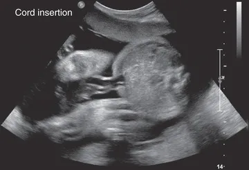
Fig. 1.8 Transverse view of the fetal abdomen and the umbilical cord insertion site showing integrity of the central abdominal wall.
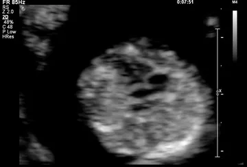
Fig. 1.9 Four-chamber view of the fetal heart at 12 weeks of gestation.
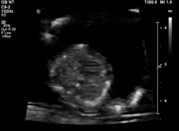
Fig. 1.10 Interventricular septum at 12 weeks of gestation.
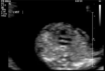
Fig. 1.11 Left ventricular outflow at 12 weeks of gestation. LVOT, Left ventricular outflow tract.
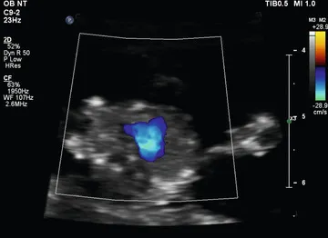
Fig. 1.12 Three-vessel view shown by color Doppler at 12 weeks of gestation.
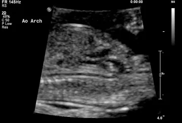
Fig. 1.13 Aortic arch (Ao Arch) at 12 weeks of gestation.
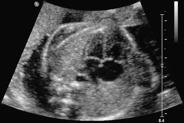
Fig. 1.14 Four-chamber view obtained with a transverse axial view through the fetal thorax. This view provides information on the size of the heart and its chambers; the pulmonary venous connections to the atrial segment; the morphology of the ventricles; the type of atrioventricular (AV) connection; and ...