
eBook - ePub
Obstetric Imaging: Fetal Diagnosis and Care E-Book
Joshua Copel, Mary E. D'Alton, Helen Feltovich, Eduard Gratacos, Anthony O. Odibo, Lawrence Platt, Boris Tutschek
This is a test
Condividi libro
- 848 pagine
- English
- ePUB (disponibile sull'app)
- Disponibile su iOS e Android
eBook - ePub
Obstetric Imaging: Fetal Diagnosis and Care E-Book
Joshua Copel, Mary E. D'Alton, Helen Feltovich, Eduard Gratacos, Anthony O. Odibo, Lawrence Platt, Boris Tutschek
Dettagli del libro
Anteprima del libro
Indice dei contenuti
Citazioni
Informazioni sul libro
Richly illustrated and comprehensive in scope, Obstetric Imaging, 2nd Edition, provides up-to-date, authoritative guidelines for more than 200 obstetric conditions and procedures, keeping you at the forefront of this fast-changing field. This highly regarded reference covers the extensive and ongoing advances in maternal and fetal imaging in a concise, newly streamlined format for quicker access to common and uncommon findings. Detailed, expert guidance, accompanied by superb, high-quality images, helps you make the most of new technologies and advances in obstetric imaging.
- Features more than 1, 350 high-quality images, including 400 in color.
- Helps you select the best imaging approaches and effectively interpret your findings with a highly templated, bulleted, at-a-glance organization.
- Reflects all the latest developments in the field, including genetics, open fetal surgery, fetal echocardiography, Zika virus, and 3D imaging, so you can provide the safest and most responsive care to both mother and fetus.
- Includes new chapters on Limbs and Bones Overview; Open Fetal Surgery; Biophysical Profile; Ultrasound Physics; Elastography; Doppler; MRI; Echogenic Bowel; Pregnancy of Unknown Location (PUL), Failed Pregnancy and Ectopic Pregnancy, Cesarean Scar Pregnancy; Cytomegalovirus (CMG), Rubella, Toxoplasmosis, Herpes, Varicella; and Congenital Syphilis; plus a new chapter on Zika Virus written by imaging experts from the "hot zone."
- Keeps you up to date with the latest developments in multimodality imaging and optimizing diagnostic accuracy from ultrasound, 3D ultrasound, Doppler, MRI, elastography, image-guided interventions, and much more.
Domande frequenti
Come faccio ad annullare l'abbonamento?
È semplicissimo: basta accedere alla sezione Account nelle Impostazioni e cliccare su "Annulla abbonamento". Dopo la cancellazione, l'abbonamento rimarrà attivo per il periodo rimanente già pagato. Per maggiori informazioni, clicca qui
È possibile scaricare libri? Se sì, come?
Al momento è possibile scaricare tramite l'app tutti i nostri libri ePub mobile-friendly. Anche la maggior parte dei nostri PDF è scaricabile e stiamo lavorando per rendere disponibile quanto prima il download di tutti gli altri file. Per maggiori informazioni, clicca qui
Che differenza c'è tra i piani?
Entrambi i piani ti danno accesso illimitato alla libreria e a tutte le funzionalità di Perlego. Le uniche differenze sono il prezzo e il periodo di abbonamento: con il piano annuale risparmierai circa il 30% rispetto a 12 rate con quello mensile.
Cos'è Perlego?
Perlego è un servizio di abbonamento a testi accademici, che ti permette di accedere a un'intera libreria online a un prezzo inferiore rispetto a quello che pagheresti per acquistare un singolo libro al mese. Con oltre 1 milione di testi suddivisi in più di 1.000 categorie, troverai sicuramente ciò che fa per te! Per maggiori informazioni, clicca qui.
Perlego supporta la sintesi vocale?
Cerca l'icona Sintesi vocale nel prossimo libro che leggerai per verificare se è possibile riprodurre l'audio. Questo strumento permette di leggere il testo a voce alta, evidenziandolo man mano che la lettura procede. Puoi aumentare o diminuire la velocità della sintesi vocale, oppure sospendere la riproduzione. Per maggiori informazioni, clicca qui.
Obstetric Imaging: Fetal Diagnosis and Care E-Book è disponibile online in formato PDF/ePub?
Sì, puoi accedere a Obstetric Imaging: Fetal Diagnosis and Care E-Book di Joshua Copel, Mary E. D'Alton, Helen Feltovich, Eduard Gratacos, Anthony O. Odibo, Lawrence Platt, Boris Tutschek in formato PDF e/o ePub, così come ad altri libri molto apprezzati nelle sezioni relative a Medizin e Medizintechnik & Zubehör. Scopri oltre 1 milione di libri disponibili nel nostro catalogo.
Informazioni
Part 1
Atlas of Selected Normal Images
1
Atlas of Selected Normal Images
Mert Ozan Bahtiyar, Carole Gravino
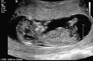
Fig. 1.1 The crown-rump length is a measurement of the length of human fetuses from the top of the crown to the bottom of the rump. It is used to estimate gestational age.
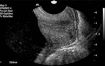
Fig. 1.2 Transvaginal ultrasound and sagittal long-axis view of the endocervical canal. Both the internal os and the external os are well visualized. The cervical length is measured from the internal os to the external os along the endocervical canal.
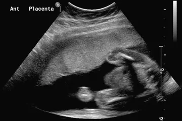
Fig. 1.3 Sagittal view of the uterus with an anterior (Ant) placenta.
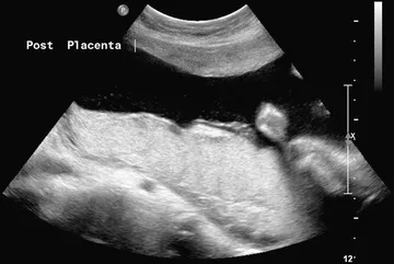
Fig. 1.4 Sagittal view of the uterus showing a posterior (Post) placenta.
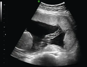
Fig. 1.5 Normal umbilical cord insertion into the placenta.
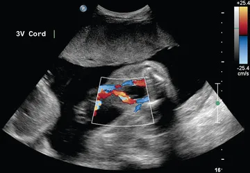
Fig. 1.6 Fetal umbilical cord insertion site. The umbilical arteries emerge caudally—originating at the iliac arteries and coursing along the margin of the urinary bladder. The umbilical vein proceeds cephalad and joins the fetal portal circulation. 3V, Three-vessel.
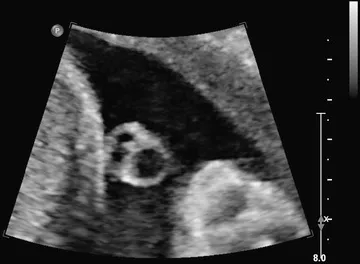
Fig. 1.7 Transverse view of the umbilical cord. The umbilical cord is composed of a vein and two smaller arteries.
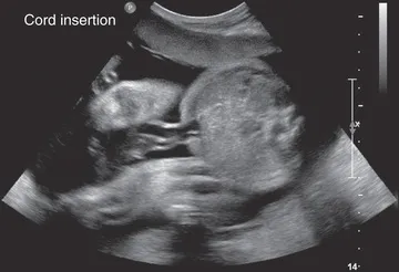
Fig. 1.8 Transverse view of the fetal abdomen and the umbilical cord insertion site showing integrity of the central abdominal wall.
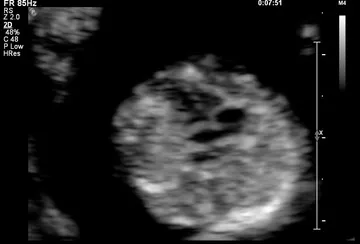
Fig. 1.9 Four-chamber view of the fetal heart at 12 weeks of gestation.
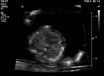
Fig. 1.10 Interventricular septum at 12 weeks of gestation.
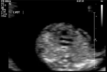
Fig. 1.11 Left ventricular outflow at 12 weeks of gestation. LVOT, Left ventricular outflow tract.
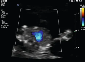
Fig. 1.12 Three-vessel view shown by color Doppler at 12 weeks of gestation.
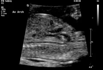
Fig. 1.13 Aortic arch (Ao Arch) at 12 weeks of gestation.
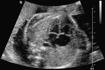
Fig. 1.14 Four-chamber view obtained with a transverse axial view through the fetal thorax. This view provides information on the size of the heart and its chambers; the pulmonary venous connections to the atrial segment; the morphology of the ventricles; the type of atrioventricular (AV) connection; and ...