
eBook - ePub
Obstetric Imaging: Fetal Diagnosis and Care E-Book
Joshua Copel, Mary E. D'Alton, Helen Feltovich, Eduard Gratacos, Anthony O. Odibo, Lawrence Platt, Boris Tutschek
This is a test
Partager le livre
- 848 pages
- English
- ePUB (adapté aux mobiles)
- Disponible sur iOS et Android
eBook - ePub
Obstetric Imaging: Fetal Diagnosis and Care E-Book
Joshua Copel, Mary E. D'Alton, Helen Feltovich, Eduard Gratacos, Anthony O. Odibo, Lawrence Platt, Boris Tutschek
Détails du livre
Aperçu du livre
Table des matières
Citations
À propos de ce livre
Richly illustrated and comprehensive in scope, Obstetric Imaging, 2nd Edition, provides up-to-date, authoritative guidelines for more than 200 obstetric conditions and procedures, keeping you at the forefront of this fast-changing field. This highly regarded reference covers the extensive and ongoing advances in maternal and fetal imaging in a concise, newly streamlined format for quicker access to common and uncommon findings. Detailed, expert guidance, accompanied by superb, high-quality images, helps you make the most of new technologies and advances in obstetric imaging.
- Features more than 1, 350 high-quality images, including 400 in color.
- Helps you select the best imaging approaches and effectively interpret your findings with a highly templated, bulleted, at-a-glance organization.
- Reflects all the latest developments in the field, including genetics, open fetal surgery, fetal echocardiography, Zika virus, and 3D imaging, so you can provide the safest and most responsive care to both mother and fetus.
- Includes new chapters on Limbs and Bones Overview; Open Fetal Surgery; Biophysical Profile; Ultrasound Physics; Elastography; Doppler; MRI; Echogenic Bowel; Pregnancy of Unknown Location (PUL), Failed Pregnancy and Ectopic Pregnancy, Cesarean Scar Pregnancy; Cytomegalovirus (CMG), Rubella, Toxoplasmosis, Herpes, Varicella; and Congenital Syphilis; plus a new chapter on Zika Virus written by imaging experts from the "hot zone."
- Keeps you up to date with the latest developments in multimodality imaging and optimizing diagnostic accuracy from ultrasound, 3D ultrasound, Doppler, MRI, elastography, image-guided interventions, and much more.
Foire aux questions
Comment puis-je résilier mon abonnement ?
Il vous suffit de vous rendre dans la section compte dans paramètres et de cliquer sur « Résilier l’abonnement ». C’est aussi simple que cela ! Une fois que vous aurez résilié votre abonnement, il restera actif pour le reste de la période pour laquelle vous avez payé. Découvrez-en plus ici.
Puis-je / comment puis-je télécharger des livres ?
Pour le moment, tous nos livres en format ePub adaptés aux mobiles peuvent être téléchargés via l’application. La plupart de nos PDF sont également disponibles en téléchargement et les autres seront téléchargeables très prochainement. Découvrez-en plus ici.
Quelle est la différence entre les formules tarifaires ?
Les deux abonnements vous donnent un accès complet à la bibliothèque et à toutes les fonctionnalités de Perlego. Les seules différences sont les tarifs ainsi que la période d’abonnement : avec l’abonnement annuel, vous économiserez environ 30 % par rapport à 12 mois d’abonnement mensuel.
Qu’est-ce que Perlego ?
Nous sommes un service d’abonnement à des ouvrages universitaires en ligne, où vous pouvez accéder à toute une bibliothèque pour un prix inférieur à celui d’un seul livre par mois. Avec plus d’un million de livres sur plus de 1 000 sujets, nous avons ce qu’il vous faut ! Découvrez-en plus ici.
Prenez-vous en charge la synthèse vocale ?
Recherchez le symbole Écouter sur votre prochain livre pour voir si vous pouvez l’écouter. L’outil Écouter lit le texte à haute voix pour vous, en surlignant le passage qui est en cours de lecture. Vous pouvez le mettre sur pause, l’accélérer ou le ralentir. Découvrez-en plus ici.
Est-ce que Obstetric Imaging: Fetal Diagnosis and Care E-Book est un PDF/ePUB en ligne ?
Oui, vous pouvez accéder à Obstetric Imaging: Fetal Diagnosis and Care E-Book par Joshua Copel, Mary E. D'Alton, Helen Feltovich, Eduard Gratacos, Anthony O. Odibo, Lawrence Platt, Boris Tutschek en format PDF et/ou ePUB ainsi qu’à d’autres livres populaires dans Medizin et Medizintechnik & Zubehör. Nous disposons de plus d’un million d’ouvrages à découvrir dans notre catalogue.
Informations
Part 1
Atlas of Selected Normal Images
1
Atlas of Selected Normal Images
Mert Ozan Bahtiyar, Carole Gravino
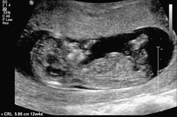
Fig. 1.1 The crown-rump length is a measurement of the length of human fetuses from the top of the crown to the bottom of the rump. It is used to estimate gestational age.
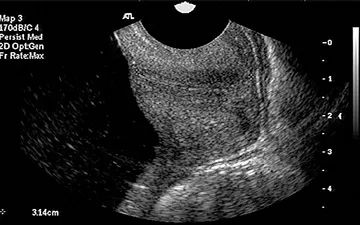
Fig. 1.2 Transvaginal ultrasound and sagittal long-axis view of the endocervical canal. Both the internal os and the external os are well visualized. The cervical length is measured from the internal os to the external os along the endocervical canal.
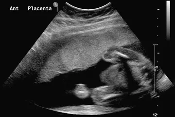
Fig. 1.3 Sagittal view of the uterus with an anterior (Ant) placenta.
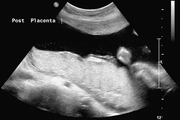
Fig. 1.4 Sagittal view of the uterus showing a posterior (Post) placenta.
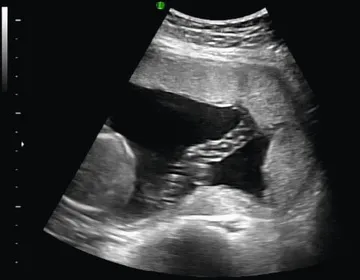
Fig. 1.5 Normal umbilical cord insertion into the placenta.
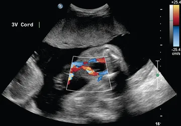
Fig. 1.6 Fetal umbilical cord insertion site. The umbilical arteries emerge caudally—originating at the iliac arteries and coursing along the margin of the urinary bladder. The umbilical vein proceeds cephalad and joins the fetal portal circulation. 3V, Three-vessel.
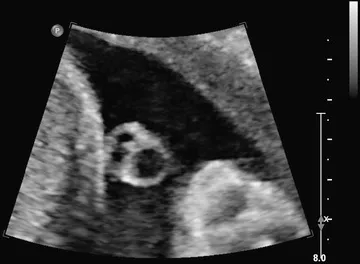
Fig. 1.7 Transverse view of the umbilical cord. The umbilical cord is composed of a vein and two smaller arteries.
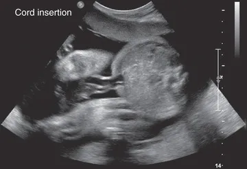
Fig. 1.8 Transverse view of the fetal abdomen and the umbilical cord insertion site showing integrity of the central abdominal wall.
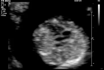
Fig. 1.9 Four-chamber view of the fetal heart at 12 weeks of gestation.
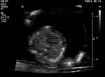
Fig. 1.10 Interventricular septum at 12 weeks of gestation.
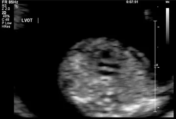
Fig. 1.11 Left ventricular outflow at 12 weeks of gestation. LVOT, Left ventricular outflow tract.
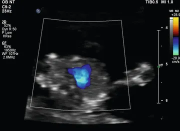
Fig. 1.12 Three-vessel view shown by color Doppler at 12 weeks of gestation.
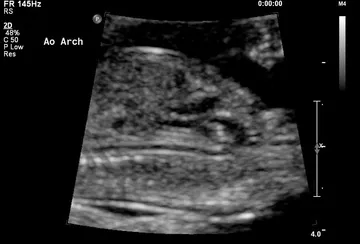
Fig. 1.13 Aortic arch (Ao Arch) at 12 weeks of gestation.
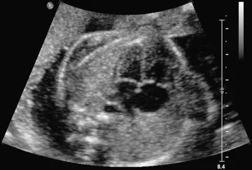
Fig. 1.14 Four-chamber view obtained with a transverse axial view through the fetal thorax. This view provides information on the size of the heart and its chambers; the pulmonary venous connections to the atrial segment; the morphology of the ventricles; the type of atrioventricular (AV) connection; and ...