
eBook - ePub
Obstetric Imaging: Fetal Diagnosis and Care E-Book
Joshua Copel, Mary E. D'Alton, Helen Feltovich, Eduard Gratacos, Anthony O. Odibo, Lawrence Platt, Boris Tutschek
This is a test
Buch teilen
- 848 Seiten
- English
- ePUB (handyfreundlich)
- Über iOS und Android verfügbar
eBook - ePub
Obstetric Imaging: Fetal Diagnosis and Care E-Book
Joshua Copel, Mary E. D'Alton, Helen Feltovich, Eduard Gratacos, Anthony O. Odibo, Lawrence Platt, Boris Tutschek
Angaben zum Buch
Buchvorschau
Inhaltsverzeichnis
Quellenangaben
Über dieses Buch
Richly illustrated and comprehensive in scope, Obstetric Imaging, 2nd Edition, provides up-to-date, authoritative guidelines for more than 200 obstetric conditions and procedures, keeping you at the forefront of this fast-changing field. This highly regarded reference covers the extensive and ongoing advances in maternal and fetal imaging in a concise, newly streamlined format for quicker access to common and uncommon findings. Detailed, expert guidance, accompanied by superb, high-quality images, helps you make the most of new technologies and advances in obstetric imaging.
- Features more than 1, 350 high-quality images, including 400 in color.
- Helps you select the best imaging approaches and effectively interpret your findings with a highly templated, bulleted, at-a-glance organization.
- Reflects all the latest developments in the field, including genetics, open fetal surgery, fetal echocardiography, Zika virus, and 3D imaging, so you can provide the safest and most responsive care to both mother and fetus.
- Includes new chapters on Limbs and Bones Overview; Open Fetal Surgery; Biophysical Profile; Ultrasound Physics; Elastography; Doppler; MRI; Echogenic Bowel; Pregnancy of Unknown Location (PUL), Failed Pregnancy and Ectopic Pregnancy, Cesarean Scar Pregnancy; Cytomegalovirus (CMG), Rubella, Toxoplasmosis, Herpes, Varicella; and Congenital Syphilis; plus a new chapter on Zika Virus written by imaging experts from the "hot zone."
- Keeps you up to date with the latest developments in multimodality imaging and optimizing diagnostic accuracy from ultrasound, 3D ultrasound, Doppler, MRI, elastography, image-guided interventions, and much more.
Häufig gestellte Fragen
Wie kann ich mein Abo kündigen?
Gehe einfach zum Kontobereich in den Einstellungen und klicke auf „Abo kündigen“ – ganz einfach. Nachdem du gekündigt hast, bleibt deine Mitgliedschaft für den verbleibenden Abozeitraum, den du bereits bezahlt hast, aktiv. Mehr Informationen hier.
(Wie) Kann ich Bücher herunterladen?
Derzeit stehen all unsere auf Mobilgeräte reagierenden ePub-Bücher zum Download über die App zur Verfügung. Die meisten unserer PDFs stehen ebenfalls zum Download bereit; wir arbeiten daran, auch die übrigen PDFs zum Download anzubieten, bei denen dies aktuell noch nicht möglich ist. Weitere Informationen hier.
Welcher Unterschied besteht bei den Preisen zwischen den Aboplänen?
Mit beiden Aboplänen erhältst du vollen Zugang zur Bibliothek und allen Funktionen von Perlego. Die einzigen Unterschiede bestehen im Preis und dem Abozeitraum: Mit dem Jahresabo sparst du auf 12 Monate gerechnet im Vergleich zum Monatsabo rund 30 %.
Was ist Perlego?
Wir sind ein Online-Abodienst für Lehrbücher, bei dem du für weniger als den Preis eines einzelnen Buches pro Monat Zugang zu einer ganzen Online-Bibliothek erhältst. Mit über 1 Million Büchern zu über 1.000 verschiedenen Themen haben wir bestimmt alles, was du brauchst! Weitere Informationen hier.
Unterstützt Perlego Text-zu-Sprache?
Achte auf das Symbol zum Vorlesen in deinem nächsten Buch, um zu sehen, ob du es dir auch anhören kannst. Bei diesem Tool wird dir Text laut vorgelesen, wobei der Text beim Vorlesen auch grafisch hervorgehoben wird. Du kannst das Vorlesen jederzeit anhalten, beschleunigen und verlangsamen. Weitere Informationen hier.
Ist Obstetric Imaging: Fetal Diagnosis and Care E-Book als Online-PDF/ePub verfügbar?
Ja, du hast Zugang zu Obstetric Imaging: Fetal Diagnosis and Care E-Book von Joshua Copel, Mary E. D'Alton, Helen Feltovich, Eduard Gratacos, Anthony O. Odibo, Lawrence Platt, Boris Tutschek im PDF- und/oder ePub-Format sowie zu anderen beliebten Büchern aus Medizin & Medizintechnik & Zubehör. Aus unserem Katalog stehen dir über 1 Million Bücher zur Verfügung.
Information
Part 1
Atlas of Selected Normal Images
1
Atlas of Selected Normal Images
Mert Ozan Bahtiyar, Carole Gravino
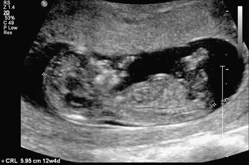
Fig. 1.1 The crown-rump length is a measurement of the length of human fetuses from the top of the crown to the bottom of the rump. It is used to estimate gestational age.
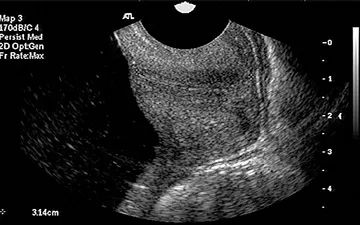
Fig. 1.2 Transvaginal ultrasound and sagittal long-axis view of the endocervical canal. Both the internal os and the external os are well visualized. The cervical length is measured from the internal os to the external os along the endocervical canal.
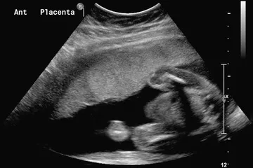
Fig. 1.3 Sagittal view of the uterus with an anterior (Ant) placenta.
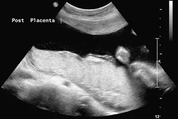
Fig. 1.4 Sagittal view of the uterus showing a posterior (Post) placenta.
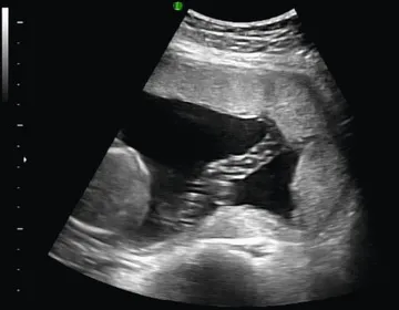
Fig. 1.5 Normal umbilical cord insertion into the placenta.
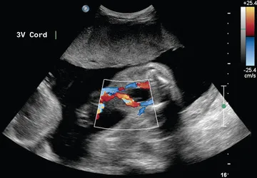
Fig. 1.6 Fetal umbilical cord insertion site. The umbilical arteries emerge caudally—originating at the iliac arteries and coursing along the margin of the urinary bladder. The umbilical vein proceeds cephalad and joins the fetal portal circulation. 3V, Three-vessel.
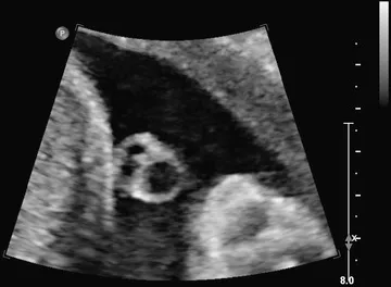
Fig. 1.7 Transverse view of the umbilical cord. The umbilical cord is composed of a vein and two smaller arteries.
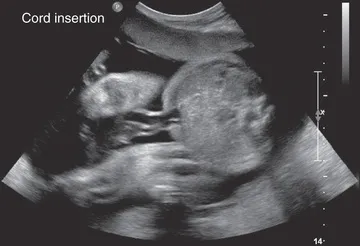
Fig. 1.8 Transverse view of the fetal abdomen and the umbilical cord insertion site showing integrity of the central abdominal wall.
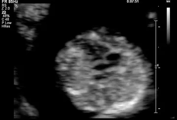
Fig. 1.9 Four-chamber view of the fetal heart at 12 weeks of gestation.
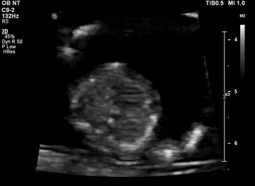
Fig. 1.10 Interventricular septum at 12 weeks of gestation.
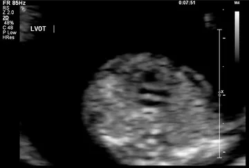
Fig. 1.11 Left ventricular outflow at 12 weeks of gestation. LVOT, Left ventricular outflow tract.
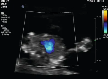
Fig. 1.12 Three-vessel view shown by color Doppler at 12 weeks of gestation.
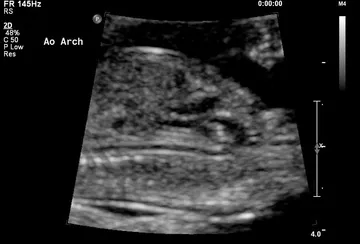
Fig. 1.13 Aortic arch (Ao Arch) at 12 weeks of gestation.
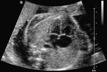
Fig. 1.14 Four-chamber view obtained with a transverse axial view through the fetal thorax. This view provides information on the size of the heart and its chambers; the pulmonary venous connections to the atrial segment; the morphology of the ventricles; the type of atrioventricular (AV) connection; and ...