Psychology
Lesioning Research
Lesioning research involves deliberately damaging or removing specific areas of the brain in order to study the resulting behavioral or cognitive changes. This method helps researchers understand the functions of different brain regions and their contributions to various psychological processes. Lesioning studies have provided valuable insights into the brain-behavior relationship and have contributed to our understanding of neurological disorders and cognitive functions.
Written by Perlego with AI-assistance
Related key terms
1 of 5
9 Key excerpts on "Lesioning Research"
 eBook - PDF
Learning and Memory
A Biological View
- Joe L. Jr. Martinez(Author)
- 2012(Publication Date)
- Academic Press(Publisher)
= ν = LESIONS This page intentionally left blank 11 INTERVENTIONAL APPROACHES TO MEMORY: LESIONS David S. Olton INTRODUCTION A lesion analysis examines the behavioral changes that follow dam-age to the brain in order to draw conclusions about the functional organization of the nervous system. This type of experimental strategy has had a profound effect on the ways in which we think about the brain, behavior, psychology, and the interrelationships among them. Historically, the lesion analysis produced all of our initial ideas about brain function (Thompson & Robinson, 1979). Everyday events like accidents, war, and disease, and experimental techniques such as abla-tion, provided direct ways of interfering with the activity of brain struc-tures before equivalent techniques were available to enhance or mea-sure brain activity. Currently, a lesion analysis is one of the three experimental strategies (stimulation and recording are the other two) that we can use to examine the functional organization of the brain, and it provides critical information that cannot be obtained from either of the other two strategies. 1 1 As discussed later (page 391), the lesion, stimulation, and recording analyses can be applied to any neural component: cells, neurotransmitters, ions, etc. Thus, this discus-sion of the lesion analysis is relevant to any intervention that disrupts normal neural activity. L E A R N I N G A N D M E M O R Y A BIOLOGICAL VIEW 379 Copyright © 1986 by Academic Press, Inc. All rights of reproduction in any form reserved. 380 DAVID S. OLTON As is the case with every approach that attempts to elucidate the interrelationships between structures and functions, the interpretation of the results from a lesion analysis requires careful attention to the assumptions and the logic of the analysis. When appropriate steps are taken, a lesion analysis provides a powerful strategy to understand the functional organization of the central nervous system. eBook - PDF
Learning and Memory
A Biological View
- Bozzano G Luisa(Author)
- 2012(Publication Date)
- Academic Press(Publisher)
= ν = LESIONS This page intentionally left blank 11 INTERVENTIONAL APPROACHES TO MEMORY: LESIONS David S. Olton INTRODUCTION A lesion analysis examines the behavioral changes that follow dam-age to the brain in order to draw conclusions about the functional organization of the nervous system. This type of experimental strategy has had a profound effect on the ways in which we think about the brain, behavior, psychology, and the interrelationships among them. Historically, the lesion analysis produced all of our initial ideas about brain function (Thompson & Robinson, 1979). Everyday events like accidents, war, and disease, and experimental techniques such as abla-tion, provided direct ways of interfering with the activity of brain struc-tures before equivalent techniques were available to enhance or mea-sure brain activity. Currently, a lesion analysis is one of the three experimental strategies (stimulation and recording are the other two) that we can use to examine the functional organization of the brain, and it provides critical information that cannot be obtained from either of the other two strategies. 1 1 As discussed later (page 391), the lesion, stimulation, and recording analyses can be applied to any neural component: cells, neurotransmitters, ions, etc. Thus, this discus-sion of the lesion analysis is relevant to any intervention that disrupts normal neural activity. LEARNING AND M E M O R Y A BIOLOGICAL VIEW 379 Copyright © 1986 by Academic Press, Inc. All rights of reproduction in any form reserved. 380 DAVID S. OLTON As is the case with every approach that attempts to elucidate the interrelationships between structures and functions, the interpretation of the results from a lesion analysis requires careful attention to the assumptions and the logic of the analysis. When appropriate steps are taken, a lesion analysis provides a powerful strategy to understand the functional organization of the central nervous system.
- Jan Bures, Olga Burešová, Joseph P. Huston(Authors)
- 2016(Publication Date)
- Elsevier(Publisher)
Chapter 4 ABLATION AND STIMULATION OF THE BRAIN 4.1. BRAIN LESIONS Elimination of a part of the brain for purposes of establishing its function or tracing its connections to other parts of the brain is still one of the most widely used approaches to the analysis of brain-behavior relations. Destruction of brain tissue is used for various purposes, and, hence, a wide arsenal of techniques is available, of which this chapter will describe the most common ones in some detail. Destructive intervention can be achieved by (a) cutting of discrete pathways or gross separation of structures with knife cuts (such as the various isole preparations and the split-brain), (b) lesioning of structures, as by passage of direct current (electrolytic lesion) or radiofrequency current (thermocoagulation) through electrodes, (c) surgical removal of tissue by knife or aspiration, (d) neurochemical lesions (long-term effects, such as depletion of catecholamines by 6-bydroxy-dopamine, or short-term effects, such as depletion of serotonin by para-chlorophenylalanine); and (e) reversible functional ablation, which can be achieved with cooling, spreading depression, local anesthesia and induction of epileptic activity. Hence, the concept of a brain lesion can be broadly defined to include destruction and separation of tissue, depletion of neuro-chemicals, especially neurotransmitters, and transient functional elimination of brain areas. The usual procedure is to interfere with brain function by one of these methods and to look for some change in behavior, whereby the behavior can be an index of sensory or motor function, or of some invoked psychological process such as learning, memory, and motivation (hunger, aggression, etc.). Hence, the methods described below can be used in conjunction with any of the behavioral experiments described earlier, which were designed to gauge these processes. No longer available |Learn more
- Jamie Ward(Author)
- 2019(Publication Date)
- Routledge(Publisher)
Chapter 5 The lesioned brain and stimulated brainDOI: 10.4324/9781351035187-5CONTENTS- Dissociations and associations in neuropsychology
- Single-Case Studies in Cognitive Neuropsychology
- Group Studies and Lesion-Deficit Analysis in Neuropsychology
- Animal Models in Neuropsychology
- Transcranial magnetic stimulation (TMS)
- Transcranial Electrical Stimulation (tES)
- Summary and key points of the chapter
- Example essay questions
- Recommended further reading
Studies of humans who have been unfortunate enough to acquire brain damage have provided a rich source of information for cognitive neuroscientists. The basic premise behind the approach is that, by studying the abnormal, it is possible to gain insights into normal function. This is a form of “reverse engineering,” in which one attempts to infer the function of a component (or region) by observing what the rest of the cognitive system can and can’t do when that component (or region) is removed. Following brain damage, it may be possible to write but not speak, or recognize objects but not faces. In this way, lesions “carve cognition at its seams” (McCarthy & Warrington, 1990 ).From a contemporary perspective, studies of the effects of brain lesions on cognition can be regarded as one example of a wider class of approaches in which brain functioning is disrupted or stimulated in some way—either in humans or animals. This stands in contrast to other methods, such as EEG and fMRI, where some aspect of brain activity is recorded and for which the relationship between brain and behavior is correlational. Figure 5.1 gives an overview of these brain manipulation methods summarized according to their approximate temporal and spatial resolutions. It includes classical pharmacological manipulations on either the whole brain or local circuits; together with invasive electrical stimulation of neurons or—in genetically modified animals—by light stimulation of neurons that contain light-sensitive proteins that excite or inhibit neural activity (opto-genetics). Primarily used in humans, there are various noninvasive brain stimulation (NIBS) techniques of which transcranial magnetic stimulation (TMS) is the most famous. TMS involves magnetic stimulation of the intact brain to produce what has been described as “virtual lesions” or “reversible lesions” (e.g.,Pascual-Leone et al., 1999). A set of newer methods (tDCS, tACS, tRNS) are based on the principle of electrical stimulation (Nitsche et al., 2008). Like TMS, transcranial electrical stimulation (tES) eBook - ePub
Perceptions and Representations
The Theoretical Bases of Brain Research and Psychology
- Keith Oatley(Author)
- 2017(Publication Date)
- Routledge(Publisher)
The above arguments do not, of course, mean that brain lesions cannot disclose important and unsuspected properties of the brain. Rather I want to suggest that they are inappropriate vehicles for asking the question of how a mechanism works. A much more appropriate target for the technique is the investigation of the brain’s resistance to damage: the surprising fact that the brain continues to go on working rather well despite enormous devastation of its parts, and despite other kinds of disruptive noise. This aspect is an apt target for the lesion technique. Clearly if the brain does continue to work quite well despite extensive damage or loss of any individual components, then this property sets it apart from almost every type of physical device with which we are familiar, in which damage to a single component typically makes the whole device defective or useless. One way of attacking this matter is to produce experimental brain lesions in order to investigate both the limits and nature of this remarkable property wherein the whole is more reliable than any of its parts, and the important work begun by Lashley on this problem will be discussed in a later chapter.A second use for the lesion is to isolate parts of the brain in the hope that the properties of these parts will be simpler than that of the whole. In a sense the neurophysiological study of a single nerve cell involves this kind of manipulation. Equally clearly the same principle is involved in studying an isolated ganglion in an invertebrate, an isolated retina, or an animal with only the spinal cord functioning and connected to the muscles. Particularly if the isolated part has some functional autonomy and is capable of giving rise to components of the behaviour of the whole organism it is entirely appropriate to suppose that studying the part may allow a simplification so that some mechanisms producing its behaviour can be more easily understood. In just the same way isolation of the teletypewriter of a computer from the main machine might be a valuable step towards investigating the particular properties of the teletypewriter. Removing all but one of its peripheral devices from a computer might be another good move, but in each case it is probably important that some normal communication channels remain. Thus though it is rather clear that study of the behaviour of animals with only the spinal cord working has led to important understanding of the nature of behaviour (e.g. Sherrington, 1906), it is by no means as clear that the study of the isolated slab of cortex (e.g. Burns, 1958) has the same kinds of potentiality for understanding how behaviour is produced, since we do not know how to communicate with it in a way that the cortex is usually communicated with, nor can we understand the significance of its responses. eBook - PDF
- Margaret W. Matlin(Author)
- 2014(Publication Date)
- Wiley(Publisher)
The area of cognitive neuroscience combines the research techniques of cognitive psychology with a variety of methods for assessing the brain’s structure and function. 2. A brain lesion refers to an area of the brain that has been destroyed by strokes and other forms of damage; it is often difficult to interpret the relationship between brain lesions and cognitive deficits. 3. In the positron emission tomography or PET-scan technique, researchers inject a small dose of radioactive chemical to see what parts of the brain are activated when a person is working on a cognitive task. 4. Functional magnetic resonance imaging (fMRI) tracks oxygen-rich blood to see what parts of the brain are active when a person is working on a cognitive task. 5. The event-related potential technique uses electrodes to track the very brief changes in the brain’s electrical activity, in response to specific stimuli. ADDITIONAL AREAS THAT CONTRIBUTE TO COGNITIVE PSYCHOLOGY Cognitive neuroscience, the topic that we have just discussed, is clearly the area that contributes most to cognitive psychology. However, we also need to consider two other topics, artificial intelligence and the broad field of cognitive science. Artificial Intelligence Artificial intelligence (AI) is a branch of computer science; it seeks to explore human cognitive processes by creating computer models that show ‘‘intelligent behavior’’ and also accomplish the same tasks that humans do (Berm ´ udez, 2010; Boden, 2004; Chrisley, 2004). Researchers in artificial intelligence have tried to explain how humans 18 CHAPTER 1 An Introduction to Cognitive Psychology recognize a face, create a mental image, and write a poem, as well as hundreds of additional cognitive accomplishments (Boden, 2004; Farah, 2004; Thagard, 2005). In this textbook, you’ll read about research on artificial intelligence in Chapter 8 (general knowledge), Chapter 9 (language comprehension), and Chapter 11 (problem solving). eBook - PDF
- Gabriel Kreiman(Author)
- 2021(Publication Date)
- Cambridge University Press(Publisher)
Moving beyond these correlations to establish causality is not a trivial matter. We will consider here two approaches that can help bring us a step closer toward understanding the relationship between neural activity in specific brain circuits and visual perception: lesions and electrical stimulation. 4.2 A Panoply of Lesion Tools to Study the Functional Role of Brain Areas in Animals Investigators take advantage of several tools to examine the effect of removing or silencing a brain area, including physical lesions, cooling experiments, pharmacological interven- tion, cell-specific ablation, molecular tools such as gene knockouts, and optogenetics. Physical lesions. One of the most widely used tools to study function in the brain has been the behavioral examination of subjects with physical lesions. It is also possible to induce lesions by injecting chemicals like neurotoxins. In non-human animals (hence- forth animals), investigators may remove specific brain areas to examine the behavioral deficits. For example, retinal ganglion cells project to the primary visual cortex (via the LGN) and to the superior colliculus. Primates with lesions to the superior colliculus are 63 4.2 A Panoply of Lesion Tools to Study the Functional Role of Brain Areas in Animals still capable of solving visual recognition tasks, whereas animals with lesions to the primary visual cortex are not. Subsequent studies examined the function of different parts of the visual cortex through lesions. Lesions to an area known as the middle temporal area (MT, also known as area V5) lead to severe impairment in the ability to discriminate motion direction, whereas lesions to the inferior temporal cortex lead to object recognition deficits. Lesion studies in animals often provide highly valuable information, but they are not always easy to interpret. First, it is challenging to make anatomically precise lesions.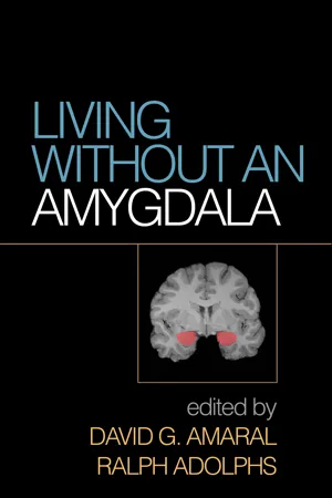 No longer available
- David G. Amaral, Ralph Adolphs, David G. Amaral, Ralph Adolphs(Authors)
- 2016(Publication Date)
- The Guilford Press(Publisher)
72 Much of the research reviewed in this book is based on the behavioral conse- quences of various forms of lesions in experimental animals and human sub- jects. A consistent theme in this research is that alterations in emotional behav- ior that are characteristic of selective amygdala lesions are far different from the behavioral outcomes of damage to the temporal neocortex in which it is embedded or to the hippocampal formation that is its neighbor. The history of how this appreciation was gained parallels the efforts of neuroscience to local- ize specific functions to anatomically defined regions of the brain. It is a story of missed opportunities, of capitalizing on unexpected findings, and of incre- mental specification of the critical locus of brain damage leading to dramatic behavioral alterations. This chapter provides a selective review of research that extends back into the late 1880s dealing with lesions and amygdala function. It also highlights the era of psychosurgery in which the results from animal lesion studies were applied to the treatment of human epileptic and psychiatric patients. While much of the history of lesions and the amygdala is based on nonprimate animal studies, this chapter deals almost entirely with studies in nonhuman primates and human subjects. Animal Studies The practice of producing lesions of the brain and observing alterations of behavior in animal models dates back to the pioneering research of the French physiologist Pierre Flourens. In studies initiated in the early 1800s, Flourens (1842) produced lesions in the brains of pigeons and rab- bits, and concluded that the cerebral cortex is responsible for sensory C H A P T E R 3 A Short History of the Lesion Technique for Probing Amygdala Function DAVID G. AMARAL The Lesion Technique for Probing Amygdala Function 73 perception, the cerebellum for motor coordination, and the medulla for heart rate, respiration and other vital bodily functions (Tizard, 1959). eBook - ePub
Cognitive Science
Contributions to Educational Practice
- Marlin L. Languis, James Buffer, Daniel Martin, Paul Naour(Authors)
- 2012(Publication Date)
- Routledge(Publisher)
Part Two Cognitive NeuroscienceResearch: Probing Substratesof Educational Problems Passage contains an image
Chapter IV
Studies of Stroke Patients Suggest a Novel Kind of Relationship Between Mood Disorder and Lesion Location*Robert G. Robinson and Thomas R. Price Introduction Behavioral Localization in the BrainThroughout much of the twentieth century, clinicians and neuroscientists have recognized that behavior involves a complex, multilevel, and integrated neuronal activation and that lesions which produce behavioral abnormalities are no more than regions that contribute to, or form part of, this integrated network leading to behavior. In spite of the recognition of this complexity of behavior, attempts to associate brain regions with particular behaviors have been very useful because they have allowed us to localize brain damage based on behavioral symptoms and to identify brain areas which are in some way related to specific behaviors. For example, Broca’s aphasia has clearly been shown to be associated with frontal dominant hemisphere injury (Benson & Geschwind, 1971), and we therefore have attributed an expressive language function to this posterior inferior region of the frontal cortex. Thus, although these kinds of brain-behavior relationships are clearly oversimplifications, clinicians and researchers have found it useful to our understanding of brain-behavior relationships to examine clinical-pathological correlations in patients with brain injury.Concepts about the relationship between brain and behavior have historically fallen into one of two diametrically opposed perspectives. The “localizers” represented by the tradition of Broca (1861) and others (Penfield & Boldrey, 1937; Benson & Geschwind, 1971) have conceived of behavioral localization as a mosaic of foci in the brain, with each foci representing discretely localized functions. This view was based on clinical-pathological correlations in which an important contribution to normal function is inferred from loss of behavior in pathological states (Wernicke, 1908) as well as the behavioral manifestations following electrical stimulation of intact tissue (Penfield & Boldrey). This idea of behavior as distributed in a “map” or “mosaic” fashion across the cortex has been a very powerful concept and has led to a basic understanding of the organization of language and motor-sensory function.
Passage contains an image
Chapter IV
Studies of Stroke Patients Suggest a Novel Kind of Relationship Between Mood Disorder and Lesion Location*Robert G. Robinson and Thomas R. Price Introduction Behavioral Localization in the BrainThroughout much of the twentieth century, clinicians and neuroscientists have recognized that behavior involves a complex, multilevel, and integrated neuronal activation and that lesions which produce behavioral abnormalities are no more than regions that contribute to, or form part of, this integrated network leading to behavior. In spite of the recognition of this complexity of behavior, attempts to associate brain regions with particular behaviors have been very useful because they have allowed us to localize brain damage based on behavioral symptoms and to identify brain areas which are in some way related to specific behaviors. For example, Broca’s aphasia has clearly been shown to be associated with frontal dominant hemisphere injury (Benson & Geschwind, 1971), and we therefore have attributed an expressive language function to this posterior inferior region of the frontal cortex. Thus, although these kinds of brain-behavior relationships are clearly oversimplifications, clinicians and researchers have found it useful to our understanding of brain-behavior relationships to examine clinical-pathological correlations in patients with brain injury.Concepts about the relationship between brain and behavior have historically fallen into one of two diametrically opposed perspectives. The “localizers” represented by the tradition of Broca (1861) and others (Penfield & Boldrey, 1937; Benson & Geschwind, 1971) have conceived of behavioral localization as a mosaic of foci in the brain, with each foci representing discretely localized functions. This view was based on clinical-pathological correlations in which an important contribution to normal function is inferred from loss of behavior in pathological states (Wernicke, 1908) as well as the behavioral manifestations following electrical stimulation of intact tissue (Penfield & Boldrey). This idea of behavior as distributed in a “map” or “mosaic” fashion across the cortex has been a very powerful concept and has led to a basic understanding of the organization of language and motor-sensory function.
Index pages curate the most relevant extracts from our library of academic textbooks. They’ve been created using an in-house natural language model (NLM), each adding context and meaning to key research topics.