- NEW! Updated content on diagnostic ultrasound ensures that you are informed about the latest developments and prepared to meet the challenges of the clinical environment.- NEW! Coverage of internal medicine includes basic knowledge about a disease process, the value of various blood tests in evaluating the disease, as well as treatment strategies.- NEW editors Rance K. Sellon and Clifford R. Berry bring a fresh focus and perspective to this classic text.- NEW! Expert Consult website includes a fully searchable eBook version of the text along with video clips demonstrating normal and abnormal conditions as they appear in ultrasound scans.- NEW! New and updated figures throughout the book demonstrate current, high-quality images from state-of-the-art equipment.- NEW contributing authors add new chapters, ensuring that this book contains current, authoritative information on the latest ultrasound techniques.
eBook - ePub
Small Animal Diagnostic Ultrasound E-Book
- 752 pages
- English
- ePUB (mobile friendly)
- Available on iOS & Android
eBook - ePub
Small Animal Diagnostic Ultrasound E-Book
About this book
Information
Topic
MedicineSubtopic
Veterinary Medicine1: Fundamentals of diagnostic ultrasound
John S. Mattoon, Clifford R. Berry
Diagnostic ultrasound uses high-frequency sound waves that are pulsed into the body, and the returning echoes are then analyzed by computer to yield high-resolution cross-sectional images of organs, tissues, and blood flow. The displayed information is a result of ultrasound interaction with tissues, which is based on the tissue’s acoustic impedance, and does not necessarily represent specific microscopic or macroscopic anatomy. Indeed, organs may appear perfectly normal on an ultrasound image in the presence of dysfunction or failure. Conversely, organs may appear abnormal on the ultrasound examination but be functioning properly. This basic tenet must be understood and respected for diagnostic ultrasound to be used properly.
High-quality ultrasound studies require a firm understanding of the important physical principles of diagnostic ultrasound. In this introductory chapter, we strive to present the necessary fundamental physical principles of ultrasound without excessive detail. In-depth sources on the subject are recommended to interested readers.1-5 These textbooks uniformly stress that image quality depends on knowledge of the interaction of sound with tissue and the skillful use of the scanner’s controls. Ultrasound examinations are highly interactive; a great deal of flexibility is often required for good images to be obtained. Accurate interpretation depends directly on the differentiation of normal and abnormal anatomy. Unlike with other imaging modalities, interpretation is best made at the time of the study. It is very difficult to render a meaningful interpretation from another sonographer’s static images or video clips.
Basic acoustic principles
Wavelength and frequency
Sound results from mechanical energy propagating through matter as a pressure wave, producing alternating compression and rarefaction bands of molecules within the conducting medium (Fig. 1.1). The distance between each band of compression or rarefaction is the sound’s wavelength (λ), the distance traveled during one cycle. Frequency is the number of times a wavelength is repeated (cycles) per second and is expressed in hertz (Hz). One cycle per second is 1 Hz; 1000 and 1 million cycles per second are 1 kilohertz (kHz) and 1 megahertz (MHz), respectively. The range of human hearing is approximately 20 to 20,000 Hz. Diagnostic ultrasound is characterized by sound waves with a frequency up to 1000 times higher than this range. Sound frequencies in the range of 2 to 15 MHz and higher are commonly used in diagnostic ultrasound examinations. Even higher frequencies (20 to 100 MHz) are used in special ocular, dermatologic, and microimaging applications.
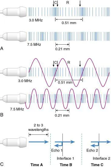
Frequencies in the millions of cycles per second have short wavelengths (submillimeter) that are essential for high-resolution imaging. The shorter the wavelength (or higher the frequency), the better the resolution. Frequency and wavelength are inversely related if the sound velocity within the medium remains constant. Because sound velocity is independent of frequency and nearly constant (1540 m/sec) in the body’s soft tissues1,5 (Table 1.1), selecting a higher frequency transducer will result in decreased wavelength of the emitted sound, providing better axial resolution (see Fig. 1.1, A, and Fig. 1.2). The relationship between velocity, frequency, and wavelength can be summarized in the following equation:

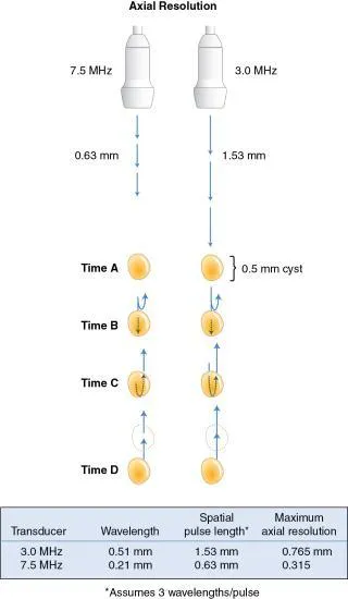
TABLE 1.1
Velocity of Sound in Body Tissues
| Tissue or Substance | Velocity (m/sec) |
|---|---|
| Air | 331 |
| Fat | 1450 |
| Water (50°C) | 1540 |
| Average soft tissue | 1540 |
| Brain | 1541 |
| Liver | 1549 |
| Kidney | 1561 |
| Blood | 1570 |
| Muscle | 1585 |
| Lens of eye | 1620 |
| Bone | 4080 |
Data from Curry TS III, Dowdey JE, Murry RC Jr. Christensen’s Physics of Diagnostic Radiology. 4th ed. Philadelphia: Lea & Febiger; 1990.
The wavelengths for commonly used ultrasound frequencies can be determined by rearranging this equation (Table 1.2).
TABLE 1.2
Commonly Used Ultrasound Frequencies *
| Frequency (MHz) | Wavelength (mm) |
|---|---|
| 2.0 | 0.77 |
| 3.0 | 0.51 |
| 5.0 | 0.31 |
| 7.5 | 0.21 |
| 10.0 | 0.15 |
*Assume velocity = 1.54 mm/µsec (1540 m/sec).
Propagation of sound
Diagnostic ultrasound uses a “pulse echo” principle in which short pulses of sound are transmitted into the body (see Fig. 1.1, C). Propagation of sound occurs in longitudinal pressure waves along the direction of particle movement as shown in Fig. 1.1. The speed of sound (propagation velocity) is affected by the physical properties of tissue, primarily the tissue’s resistance to compression, which depends on tissue density and elasticity (stiffness). Propagation velocity is increased in stiff tissues and decreased in tissues of high density. Fortunately, the propagation velocities in the soft tissues of the body are very similar, and it is therefore assumed that the average velocity of diagnostic ultrasound is 1540 m/sec.
The ultrasound transducer both sends pulses into the tissue (1% of the time) and receives the returning echoes (99% of the time). The assumption of a constant propagation velocity (1540 m/sec) is fundamental to how the ultrasound machine calculates the distance (or depth) of a reflecting surface. Suppose it takes 0.126 msec from the time of pulse until the return of the echo. The depth of the reflective surface would be calculated as follows:

This value must be divided by 2 to account for the round trip to and from the reflector, so the depth of the reflective surface equals 9.70 cm.
It should be intuitive then that if the sound travels through fatty tissue at 1450 m/sec, the reflector depth will be erroneously calculated as greater (or deeper) than it actually is. This is termed the speed propagation error and is discussed and illustrated further in later sections that focus on artifacts.
Further, when the ultrasound beam encounters gas (331 m/sec) or bone (4080 m/sec), marked velocity differences in these media result in high reflection and improper echo interpretation with characteristic reverberation and shadowing artifacts (see later sections on artifacts) (Fig....
Table of contents
- Cover image
- Title page
- Table of Contents
- Copyright
- Dedication
- Contributors
- Preface
- Acknowledgments
- Video contents
- List of Tables
- List of Illustrations
- 1. Fundamentals of diagnostic ultrasound
- 2. Ultrasound-guided aspiration and biopsy procedures
- 3. Point-of-care ultrasound
- 4. Abdominal ultrasound scanning techniques
- 5. Eye
- 6. Neck
- 7. Thorax
- 8. Echocardiography
- 9. Liver
- 10. Spleen
- 11. Pancreas
- 12. Gastrointestinal tract
- 13. Peritoneal fluid, lymph nodes, masses, peritoneal cavity, and great vessel thrombosis
- 14. Musculoskeletal system
- 15. Adrenal glands
- 16. Urinary tract
- 17. Prostate and testes
- 18. Ovaries and uterus
- Index
Trusted by 375,005 students
Access to over 1.5 million titles for a fair monthly price.
Study more efficiently using our study tools.
Frequently asked questions
Yes, you can cancel anytime from the Subscription tab in your account settings on the Perlego website. Your subscription will stay active until the end of your current billing period. Learn how to cancel your subscription
No, books cannot be downloaded as external files, such as PDFs, for use outside of Perlego. However, you can download books within the Perlego app for offline reading on mobile or tablet. Learn how to download books offline
Perlego offers two plans: Essential and Complete
- Essential is ideal for learners and professionals who enjoy exploring a wide range of subjects. Access the Essential Library with 800,000+ trusted titles and best-sellers across business, personal growth, and the humanities. Includes unlimited reading time and Standard Read Aloud voice.
- Complete: Perfect for advanced learners and researchers needing full, unrestricted access. Unlock 1.5M+ books across hundreds of subjects, including academic and specialized titles. The Complete Plan also includes advanced features like Premium Read Aloud and Research Assistant.
We are an online textbook subscription service, where you can get access to an entire online library for less than the price of a single book per month. With over 1.5 million books across 990+ topics, we’ve got you covered! Learn about our mission
Look out for the read-aloud symbol on your next book to see if you can listen to it. The read-aloud tool reads text aloud for you, highlighting the text as it is being read. You can pause it, speed it up and slow it down. Learn more about Read Aloud
Yes! You can use the Perlego app on both iOS and Android devices to read anytime, anywhere — even offline. Perfect for commutes or when you’re on the go.
Please note we cannot support devices running on iOS 13 and Android 7 or earlier. Learn more about using the app
Please note we cannot support devices running on iOS 13 and Android 7 or earlier. Learn more about using the app
Yes, you can access Small Animal Diagnostic Ultrasound E-Book by John S. Mattoon,Rance K. Sellon,Clifford Rudd Berry in PDF and/or ePUB format, as well as other popular books in Medicine & Veterinary Medicine. We have over 1.5 million books available in our catalogue for you to explore.