Biological Sciences
Chytridiomycota
Chytridiomycota is a phylum of fungi that are characterized by their unique flagellated spores, distinguishing them from other fungal groups. They are primarily aquatic and can be found in various freshwater and marine environments. Some species within this phylum are known to be pathogenic to amphibians, making them a subject of interest in conservation biology and disease ecology.
Written by Perlego with AI-assistance
Related key terms
1 of 5
12 Key excerpts on "Chytridiomycota"
 eBook - PDF
- John Webster, Roland Weber(Authors)
- 2007(Publication Date)
- Cambridge University Press(Publisher)
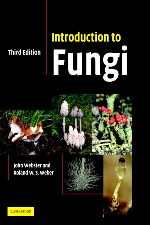
6 Chytridiomycota 6.1 Introduction The phylum Chytridiomycota comprises over 900 species in five orders (D. J. S. Barr, 2001; Kirk et al ., 2001). Fungi included here are colloquially called ‘chytrids’. Most chytrids grow aerobically in soil, mud or water and reproduce by zoospores with a single posterior flagellum of the whiplash type, although the zoospores of some members of the Neocallimastigales are multiflagellate. Some species inhabit estuaries and others the sea. Sparrow (1960) has given an extensive account of aquatic forms, Karling (1977) a compendium of illustrations, and Powell (1993) has provided examples of the importance of the group. Many members are saprotrophs, utilizing cellulose, chitin, keratin, etc., from decaying plant and animal debris in soil and mud, whilst species of Caulochytrium grow as mycoparasites on the mycelium and conidia of terrestrial fungi (Voos, 1969). Saprotrophs can be obtained in crude culture by floating baits such as cellophane, hair, shrimp exoskeleton, boiled grass leaves and pollen on the surface of water overlying samples of soil, mud or pieces of aquatic plant material (Sparrow, 1960; Stevens, 1974; Willoughby, 2001). From such crude material, pure cultures may be prepared by streaking or pipetting zoospores onto agar containing suitable nutrients and antibiotics to limit con-tamination from bacteria. The growth and appearance of chytrids in pure culture is variable and often differs significantly from their natural habit. This has led to problems in classification systems based on thallus morphology (Barr, 1990, 2001). The availability of cultures has, however, facilitated studies on chytrid nutrition and physiology (Gleason, 1976). Some chytrids are biotrophic parasites of filamentous algae and diatoms and may severely deplete the population of freshwater phyto-plankton (see p. 139). eBook - ePub
- Thomas N Taylor, Michael Krings, Edith L. Taylor(Authors)
- 2014(Publication Date)
- Academic Press(Publisher)
Letcher et al., 2005 ).Figure 4.7 John S. Karling (left) and James P. Braselton.Figure 4.8 Donald J. S. Barr.Figure 4.9 Martha J. Powell.Today there are more than 1250 named species of chytrids and the group is global in distribution (Shearer et al., 2007 ). The Chytridiomycota have long been regarded as the sister group to the remaining fungal phyla and were once thought of as monophyletic, based on features of the life cycle and characters of the zoospores. They have a long and interesting history regarding their placement on the tree of life. At one time they were considered true fungi, based on the chitin in their cell walls (Bartnicki-Garcia, 1970 ), but this interpretation was later modified and the group was thought to represent a transitional group between protists and Fungi because of the presence of motile zoospores (Barr, 1990 ). Subsequent phylogenetic studies based on ribosomal RNA confirmed the group as true fungi and they are now regarded as basal among the Fungi (James et al., 2000 ). Although earlier studies based on zoospore ultrastructure and certain morphologic features suggested the group to be monophyletic, some more recent research suggests that the chytrids are polyphyletic or paraphyletic (e.g. Tanabe et al., 2005 ; James et al., 2006a ). In a comprehensive phylogenetic classification of fungi based on multilocus datasets, Hibbett et al. (2007) elevate the Blastocladiomycota and Neocallimastigomycota, two groups previously included within the Chytridiomycota, to the level of phyla (Figure 4.10 ). Based on these studies and those involving zoospore ultrastructure and morphology, some authorities now subdivide the remaining Chytridiomycota into six orders: Chytridiales, Cladochytriales, Lobulomycetales, Rhizophlyctidales, Rhizophydiales, Spizellomycetales; and two additional clades (Longcore, 2009 ). There will ultimately be additional revisions to fungal classifications as tools and analytical techniques continue to improve, especially in these basal fungal lineages (Hibbett et al., 2007 ), and the use of molecular tools indicates that chytrids are far more diverse than previously understood (Letcher et al., 2006 , 2008a , b ; Marano et al., 2012 eBook - ePub
Current Developments in Biotechnology and Bioengineering
Filamentous Fungi Biorefinery
- Mohammad Taherzadeh, Jorge Ferreira, Ashok Pandey(Authors)
- 2022(Publication Date)
- Elsevier(Publisher)
Members of the zoosporic true fungi are Blastocladiomycota, Chytridiomycota, and Neocallimastigomycota. Taxonomical identification of Chytrid considerably relies on a combination of ultrastructure and molecular data because majority of Chytridiomycota species are aquatic habitants and have rarely been cultured for studying and taxonomic purposes. Most were classified as “uncultured” and thus any speculations on a chytrid's ecological role based on the literature is shifty due to the difficulty of assigning names to environmental sequences that match poorly to the databases. Chytrids are one of the early diverging fungal lineages and is considered to be one of the most ancestral groups of fungi. Their membership in the fungal kingdom is demonstrated by the presence of chitin cell walls, a posterior whiplash flagellum, absorptive nutrition, use of glycogen as an energy storage compound, synthesis of lysine by the α-amino adipic acid (AAA) pathway, Golgi apparatus with stacked cisternae, and nuclear envelope fenestrated at poles during mitosis.General morphological features of chytrids.- 1. Motile asexual zoospores (with a single posterior flagellum).
- 2. Both a kinetosome and nonfunctional centriole.
- 3. Nine flagellar props and a microbody-lipid globule complex in zoosporangia.
- 4. Presence of a thallus that may be holocarpic (where thallus is involved in formation of the sporangium) or eucarpic (only part of the thallus is converted into the fruiting body), monocentric, polycentric, unicellular, or filamentous.
- 5. Sexual reproduction accompanies zygotic meiosis, that produces motile sexual zoogametes; sexual reproduction not oogamous.
- 6. Asexual reproduction by zoospores bearing a single posteriorly directed flagellum.
- 7. Zoospores containing a kinetosome and a nonflagellated centriole.
- 8. Thallus monocentric or rhizomycelial polycentric.
Chytrids are important pathogens of plants (e.g., Synchitrium), animals (e.g., Batrachochytrium), parasites to several groups of algae (e.g., Chytridium, Dinomyces). Chytrids are efficient decomposers of highly recalcitrant organic matter, such as pollen (e.g., Spizellomyces, Rhyzophidium), cellulose (e.g., Rhizophlyctis), arthropod exoskeletons, and fungal spores. Due to its parasitoidism, existence chytrids are unculturable resulting in very limited sequence information on zoosporic fungi, which poses challenges in obtaining a robust chytrid tree of life (Grossart et al., 2016 ). A well-resolved phylogenetic backbone of the fungal tree of life is required to describe how the fungal nutritional toolkit has evolved over a billion years. However, with modern day tools and technology this situation is changing. With the use of single-cell-based techniques genomic environmental sampling is steadily increasing. Single-cell genomics method was used for the first time to uncultured mycoparasitic EDF from the Cryptomycota, Chytridiomycota, and Zoopagomycota (Ahrendt et al., 2018 ). The Chytridiomycota have attracted the attention of mycologists due to their potential for negatively affecting phytoplankton communities, and altering phytoplankton dynamics throughout the season. Kagami et al. showed that Chytridiomycota effectively channeled organic matter and energy to higher trophic levels, a mechanism which has been termed the “mycoloop” (Kagami et al., 2014 eBook - PDF
Handbook of Soil Sciences
Properties and Processes, Second Edition
- Pan Ming Huang, Yuncong Li, Malcolm E. Sumner, Pan Ming Huang, Yuncong Li, Malcolm E. Sumner(Authors)
- 2011(Publication Date)
- CRC Press(Publisher)
The soil chytrids possess motile zoospores pow-ered by a single, posterior flagellum, and are thought to be close to the common ancestor of fungi and animals. Phylogenetic analyses that have included more than one representative of each of the chytrids and zygomycetes have indicated that the chytrids may form a monophyletic group, splitting the zygomycetes (Van de Peer and De Wachter, 1997) or that both groups may be poly-phyletic and interspersed one among the other (Nagahama et al., 1995; James et al., 2006b). Together, the chytrids and zygomyce-tes differ considerably from the Ascomycota and Basidiomycota in that they are typically coenocytic, with multiple geneti-cally different nuclei per cell, whereas the Ascomycota and Basidiomycota typically have uninucleate primary mycelia and binucleate (dikaryotic) secondary mycelia. Many of the approxi-mately 700 described species of chytrids (Kirk et al., 2008) occur in freshwater or marine environments, and I know of no esti-mate of the number of species occurring in soil. 24.3.2.5 Kingdom Fungi, Phylum Zygomycota Soil-inhabiting members of the Zygomycota belong in two groups of very different morphology and ecology: the predomi-nantly saprobic zygomycetes (Mucorales and relatives) and the Zoopagales, Kickxellales, and Entomophthorales, which are parasitic on insects and other invertebrates, fungi, and humans (O’Donnell, 1979; Humber, 1989; Alexopoulos et al., 1996; Benny et al., 2001; White et al., 2006; Kirk et al., 2008). A third group formerly treated as the Glomerales (misspelled Glomales) has since been shown to represent an independent phylum, Glomeromycota (Schüßler et al., 2001; see later). As mentioned above, recent phylogenetic analyses that have included multiple examples of both chytrids and Zygomycota suggest that neither forms a monophyletic group. eBook - PDF
Marine Fungi
and Fungal-like Organisms
- E. B. Gareth Jones, Ka-Lai Pang, E. B. Gareth Jones, Ka-Lai Pang(Authors)
- 2012(Publication Date)
- De Gruyter(Publisher)
The ecology of chytrids in aquatic ecosys-tems: roles in food web dynamics. Fungal Biol Rev. 2008;2:17–25. Gleason FH, Marano AM, Johnson P, Martin WW. Blastocladian parasites of invertebrates. Fungal Biol Rev. 2010;24:56–67. Glockling SL, Beakes GW. An ultrastructural study of development and reproduction in the nema-tode parasite, Myzocytiopsis vermicola. Mycologia 2006;98:7–21. Glücksman E, Bell T, Griffiths RI, Bass D. Closely related protist strains have different grazing impacts on natural bacterial communities. Environ Microbiol. 2010;12:3105–3113. Heger TJ, Mitchell EAD, Todorov M, Golemansky V, Lara E, Leander BS, Pawlowski J. Molecular phylogeny of euglyphid testate amoebae (Cercozoa: Euglyphida) suggests transitions between marine supralittoral and freshwater/terrestrial environments are infrequent. Mol Phylogenet Evol. 2010;55:113–122. Honda D, Yokochi T, Nakahara T, Raghukumar S, Nakagiri A, Schauman K, Higashihara T. Molec-ular phylogeny of labyrinthulids and thraustochytrids based on the sequencing of 18S ribosomal RNA gene. J Eukaryot Microbiol. 1999;46:637–647. James TY, Letcher PM, Longcore JE, Mozley-Standridge SE, Porter D, Powell MJ, Griffith GW, Vilgalys R. A molecular phylogeny of the flagellated fungi (Chytridiomycota) and description of a new phylum (Blastocladiomycota). Mycologia 2006;98:860–871. Jones MDM, Forn I, Gadelha C, Egan MJ, Bass D, Massana R, Richards TA. Discovery of novel intermediate forms redefines the fungal tree of life. Nature 2011;474:200–203. Kagami M, de Bruin A, Ibelings BW, van Donk E. Parasitic chytrids: their effects on phytoplankton communities and food-web dynamics. Hydrobiol. 2007;578:113–129. Kagami M, Helmsing NR, van Donk E. Parasitic chytrids could promote copepod survival by mediating material transfer from inedible diatoms. Hydrobiol. 2011;659:49–54. Kim H, Gandhi SR, Moreau RA, Weete DJ. Lipids of Haliphthoros philippinensis : an oomycetous marine microbe. J Amer Oil Chem Soc. 1998;75:1657–1665. eBook - PDF
- Lawrence A. Lacey(Author)
- 1997(Publication Date)
- Academic Press(Publisher)
CHAPTER V-4 Fungi: Oomycetes and Chytri di omyc ete s JAMES L. KERWIN & ERIN E. PETERSEN Botany Department, University of Washington, Seattle, Washington 98195 USA. 1 INTRODUCTION - PHYLOGENY AND LIFE CYCLES Aquatic fungi, formerly grouped together as Phycomycetes, are a diverse group of organisms characterized by a motile, flagellated zoospore at some stage of their life cycle (Sparrow, 1960). It is now recognized that there are phylogenetically distinct groups within this artificial assemblage. This chapter will deal primarily with two genera of entomopathogenic organisms, Lagenidium (Oomycetes: Lagenidiales) and Coelomomyces spp. (Chytridiomycetes: Blastocladiales). These are two very different groups of organisms, with one common feature: Lagenidium giganteum, the species discussed in detail here, and Coelomomyces spp. are primarily parasites of mosquito larvae. Because they are parasites of these medically important arthropods, there has been much interest for the last two decades in developing them for use in operational mosquito control. MANUALOF TECHNIQUESIN INSECTPATHOLOGY ISBN 0-12-432555-6 A Lagenidium giganteum The Oomycetes are a group of organisms whose phy- logenetic relationship to fungi and other taxonomic groups has been the subject of controversy for many years (Copeland, 1956; Barr, 1992). Although there are dissenting opinions, the prevailing view is that Oomycetes are related to heterokont algae, and are placed in the kingdom Chromista, which includes diatoms, the brown algae and all protists with chloro- plast endoplasmic reticulum or tubular ciliary mastigonemes. Monographs describing the relation- ship of the Lagenidiales to related taxa by Karling (1981) and a revision of earlier views (Dick, 1996) are available. Lagenidium giganteum is a robust, fast-growing facultative parasite which can be grown on a variety of undefined media (Domnas et al., 1982; Kerwin et al., 1986). eBook - ePub
- P. J. Quinn, B. K. Markey, F. C. Leonard, P. Hartigan, S. Fanning, E. S. Fitzpatrick(Authors)
- 2011(Publication Date)
- Wiley-Blackwell(Publisher)
Mucormycotina in the future as the term is still in use.Key points- Eukaryotic, non-photosynthetic microorganisms in the kingdom Fungi
- Widely distributed in the environment
- Cell walls contain chitin and other polysaccharides
- Heterotrophs; produce exoenzymes and obtain nutrients by absorption
- Branching hyphae and unicellular yeasts are the two major forms
- Reproduce both sexually and asexually with the production of spores
- Grow aerobically at 25°C; some moulds are strict aerobes
- Tolerate high osmotic pressures and low pH values; grow on Sabouraud dextrose agar, pH 5.5
- Resistant to antimicrobial drugs which are effective against bacteria
- Majority are saprophytes; some cause opportunistic infections
- Dermatophytes are pathogens that cause ringworm in animals and humans
Traditional classification of the fungi relies heavily on morphology and sexual reproduction. The form of a fungal species during its sexually reproductive life cycle is termed its teleomorph, while its asexual form is referred to as its anamorph. The preferred term for fungi that lack a meiotic stage is mitosporic fungi. About a fifth of all fungi, including many Aspergillus, Malassezia, Penicillium and Coccidioides species, have no known sexual stage. Formerly these fungi were placed in a heterogeneous group called the Class Deuteromycota or Fungi Imperfecti. Molecular methods are increasingly being used to produce phylogenetic trees which demonstrate evolutionary relationships and to assign fungal species to their appropriate grouping, even where there is no known sexual form. Such methods usually involve comparisons of the nucleotide sequences of highly conserved ribosomal RNA, especially the small (18S) subunit (SSU) and the large (26S) subunit (LSU) of ribosomal DNA. Many fungi formerly assigned to the Fungi Imperfecti have been transferred to the Ascomycota . A dual naming system has been used for many years with separate teleomorphic and anamorphic names. This system arose because many fungi were identified before their sexual reproductive mode was recognized. In many instances the anamorphic name is better known because it is the asexual form which is associated with disease production. This is illustrated by the dermatophyte Microsporum canis whose teleomorph name is Arthroderma otae . It is expected that, with advances in diagnostic molecular methods, the dual naming system will eventually become unnecessary. Fungi of veterinary importance are found in the three phyla Ascomycota, Basidiomycota and Zygomycota . Members of the Chytridiomycota cause skin infections in frogs which interfere with their ability to respire through their skin (Rosenblum et al ., 2010). Chytridiomycosis, first reported in Australia, has subsequently been reported in many continents and has resulted in high mortality rates in amphibians in California, Central America and regions of South America. The glomeromycetes, which are the smallest group of fungi, are of major ecological importance as plant symbionts which form arbuscular mycorrhizal associations with plant roots. The phylum Blastocladiomycota eBook - PDF
Reconstructing the Tree of Life
Taxonomy and Systematics of Species Rich Taxa
- Trevor R. Hodkinson, John A.N. Parnell(Authors)
- 2006(Publication Date)
- CRC Press(Publisher)
There are two basal evolutionary branches (sister groups to the rest of Hymenomycetes), one leading to Tremel-lales (jelly fungi) and the other to Dacrymycetales, Auriculariales (tree ear fungi), Agaricales (mushrooms) and Aphyllophorales (shelf fungi). Agaricales contains many names that have been known since humans started to collect mushrooms. Amanita (Figure 15.1e) is a genus commonly associated with mushroom poisoning, whilst Agaricus bitorquis (button mushroom), Flammulina velutipes (Enokitake), Lentinula edodes (shiitake), and Pleurotus ostreatus (oyster mushroom) are widely known as food. 15.1.4 C HYTRIDIOMYCOTA The Chytridiomycota is the only fungal phylum that produces motile zoospores and requires water for dispersal (Figure 15.1f–g). They have been classified in the Protista and Protoctista 38,39 , but based on SSU rDNA sequence they were recently included in the kingdom Fungi 40 . Chytridiomycota are probably a very ancient group, with extant forms possibly having changed little since the early periods of eukaryotic evolution 41 . They are commonly found in lakes, streams, ponds, roadside ditches and coastal marine environments, as well as in soil. As members of terrestrial and aquatic microbial communities, chytrids play an important ecological role in decomposition of chitin, cellulose, keratin and hemicellulose 42 . Notable plant pathogenic species include Synchytrium endobioticum (potato black wart), Physoderma maydis (corn brown spot) and Urophlytis alfalfae (alfalfa crown wart). As a representative of lower fungi, Allomyces macrogynus has become fashionable in molecular biology for the comparative study of the primitive genetic features with other higher fungi 43–45 . 15.1.5 Z YGOMYCOTA The phylum Zygomycota (Gr. zygos, yoke of marriage; mykes, fungi) is principally characterised by the presence of nonseptate (coenocytic) mycelium and the production of dark, thick-walled, ornamented sexual spores, called zygospores (Figure 15.1h–i). eBook - ePub
Evolution
The Origins and Mechanisms of Diversity
- Jonathan Bard(Author)
- 2021(Publication Date)
- CRC Press(Publisher)
Laraba et al., 2020) .(a): The natural flowers of the grass, Xyris surinamensis, on its cone-like spike. (b): The pseudoflowers of F. xyrophilume, a fungus that has infected the grass (scale bar: 5 mm).Figure10.8(From Laraba I et al. (2020) F. xyrophilum, sp. Nov., a member of the Fusarium fujikuroi species complex recovered from pseudoflowers on yellow-eyed grass (Xyris spp.) from Guyana. Mycologia; 112:39–51.)Chytridomycota are aquatic and can be found in ponds. It is probably for this reason that they have zoospores, motile spores with a flagellum; they are thus the only fungal taxon that maintains the ancestral state (Liu et al., 2006) . The Cryptomycota are unusual: the very few taxa are the only fungi that lack chitin in their cell membrane, although there is some in their spores (James & Berbee, 2012) . Recent phylogenetic analysis places them very closely related to the microsporidia (Bass et al., 2018).Multicellular fungi are very widely spread today, with fungal spores being almost ubiquitous: they are present in every land environment and there are even marine taxa (Sun et al., 2019) . One reason for their success is that fungi have not only evolved to survive alone in a very wide range of habitats but also to live socially with other fungi (Fig. 10.9 ). Another reason is that they have also adapted to a shared life with organisms from other phyla as commensals (having little effect on them), as parasites and as symbionts (the classic example here is lichen, where the partner taxon may be an alga or a cyanobacterium - see Chapter 19) . Many plants and fungi now have a symbiotic existence and the region of interaction between them is known as a mycorrhiza. As to the relative success of fungi, Bar-On et al. (2018) eBook - PDF
- Rodney P. Anderson, Linda Young, Kim R. Finer(Authors)
- 2020(Publication Date)
- Wiley(Publisher)
The Chytridiomycota reproduce with flagellated zoospores. Lichens exhibit mutualistic symbiotic relationships between fungi and photosynthetic organisms, such as green algae. • Numerous fungi are pathogenic to humans. Pathogenic fungi are often grouped based upon tissues targeted and may be clas- sified as superficial, cutaneous, subcutaneous, systemic, or opportunistic mycoses. Rather than causing infection, some fungi produce mycotoxins (ergot and aflatoxin), which cause organ damage in humans. 7.4 The Helminths 159 • The long, thin, bilaterally symmetric body plan of helminths is evolutionarily ancient and highly successful in terms of both species diversity and abundance. In addition to many free-living species, numerous worms have adapted to life as human para- sites and represent significant global morbidity. • Cestodes (tapeworms) and trematodes (flukes) are two classes of parasitic worms in the phylum Platyhelminthes (flatworms). Annelids (earthworms and leeches) include seg- mented worms with more highly developed organ systems than those of flatworms. The most abundant worms are the nema- todes (roundworms). Members of this phylum have proved ben- eficial as a model research organism and harmful as both human and agricultural pathogens. • Tapeworms, pinworms, and hookworms are common worms that infect the gastrointestinal system, causing significant global morbidity. Life-threatening systemic infections such as schistosomiasis and sight-threatening infections such as river blindness and loa loa are also caused by helminthic pathogens. 7.5 The Arthropods 163 • Based on species diversity and abundance, arthropods are the most successful group of animals. They possess a jointed exo- skeleton of protein and chitin to provide support and protec- tion and have evolved complex organ systems. Two arthropod subphyla contain members that either act as human pathogens or serve as vectors of transmission.
- Imran Ul Haq, Siddra Ijaz, Iqrar Ahmad Khan, Imran Ul Haq, Siddra Ijaz, Iqrar Ahmad Khan(Authors)
- 2022(Publication Date)
- CRC Press(Publisher)
2 An Insight Into Fungal Biology Imran Ul Haq, Nabeeha Aslam Khan and Muhammad Kaleem SarwarDOI: 10.1201/9781003162742-2CONTENTS
- Introduction to Fungi
- Physiology and Biochemical Genetics
- Nutritional Requirement, Uptake, Assimilation, and Growth
- Spore Germination and Penetration
- Biotrophic Phase
- Necrotrophic Phase
- Sporulation
- Fungal Metabolism
- Nitrogen Metabolism
- Carbon Catabolism
- Cell Wall Formation
- Composition
- Reproduction
- Vegetative Mode of Reproduction
- Fragmentation
- Budding
- Fission
- Rhizornorphs
- Oidia
- Sclerotia
- Chlamydospores
- Asexual Reproduction
- Sporangiospores
- Conidiospores
- Zoospores
- Sexual Reproduction
- Plasmogamy
- Karyogamy
- Meiosis
- Planogametic Copulation
- Gametangial Contact
- Gametangial Copulation
- Somatogamy
- Specialization
- Morphogenesis
- Chitosan
- Pathogenesis
- Host Specificity
- Defense Systems
- Regulation of Pathogenesis
- Invasion of Pathogen
- Colonization and Alteration of Host Physiology
- Reproduction
- Sexual Reproduction and Mating Systems
- Mating Systems
- Homothallism and Heterothallism
- Response to Stress and Sensing
- Quorum Sensing
- References
Introduction to Fungi
Fungi are the second largest group of living organisms, with 51,000 genera and more than 70,000 known species (Blackwell, 2011 ; Hawksworth, D.L., 2012 ). Fungi are eukaryotic, multicellular heterotrophs. Fungi are organisms with chitin in their cell walls as the main structural component. The other components of their cell wall are mainly glucan, mannan, and chitosan. About 22–44% of the fungal cell wall is chitin as distinguished from the cell wall of plants (Khale and Deshpande, 1992 ; Muzzarelli et al., 1994 ; Kirk et al., 2008 ), and fungi exhibit the absorptive mode of nutrition (as distinguished from animals). The body of fungi is filamentous, called “mycelium.” The mycelium is composed of tubular cells known as “hyphae” (aseptate or septate). The body of the fungus is called “thallus.” Some fungi are biotrophs (obtain nutrition from living cells), some are saprotrophs (get nutrition from the dead host), and some are necrotrophs (attack and kill the host, then obtain nutrients). They are parasites, decomposers, and also exhibit symbiosis. The true fungi are divided into four main phyla: Basidiomycota, Ascomycota, Zygomycota, and Chytridiomycota (Alexopoulos et al., 1996 ; Webster and Weber, 2007 eBook - ePub
Wood Microbiology
Decay and Its Prevention
- Robert A. Zabel, Jeffrey J. Morrell(Authors)
- 2020(Publication Date)
- Academic Press(Publisher)
Chapter ThreeThe characteristics and classification of fungi and bacteria
Abstract
This chapter discusses the classification of fungi and bacteria in related to the classic approach using morphology vs more recent developments using DNA sequencing methods.Keywords
Fungi; Bacteria; White rot; Brown rotFungi and bacteria are the principal microorganisms that invade wood during its growth, processing, storage, or use, causing decay or other property changes.Contents
Outline- Fungi in relation to other life forms 56
- Bacteria 58
- Fungi 59
- Macroscopic appearances of fungi 60
- Microscopic features of fungi 62
- Hyphal wall structure 64
- Fungal ultrastructure 67
- Specialized hyphae 68
- Cultural characteristics 70
- DNA sequencing methods 71
- Classic fungal identification by culturing 71
- Fungal reproduction 73
- Life cycles 76
- Reproductive capacity 79
- Fungal variability 79
- Growth requirements 81
- A classification of fungi 81
- Basidiomycota 84
- Agaricales 85
- Hymenochaetales 86
- Gloeophyllales 86
- Boletales 86
- Polyporales 87
- Russulales 87
- Cantharellales 88
- Deuteromycetes or fungi imperfectii 88
- A classification of bacteria 89
- Cytophagales 90
- Gram-negative facultatively anaerobic rods 90
- Gram-negative, aerobic rods, and cocci 91
- Endospore-forming rods and cocci 91
- Actinobacteria 91
- The roles of fungi and bacteria in ecosystems and human affairs 92
- Summary 93
- References 95
- Further reading 97
An understanding of the decay process and its prevention or control depends, in part, on understanding the features and capabilities of these decay agents. This chapter emphasizes the unique nature of fungi and their relationship to the other major life forms. It reviews fungal structures, growth patterns, life cycles, reproductive modes, and variable features of fungi, placing emphasis on the wood-inhabitors. Classification systems are presented to facilitate the taxonomic placement and recognition of some of the major wood-inhabiting microorganisms. Since bacteria cause only minor damage to structural wood in most environments, they are only briefly covered.
Index pages curate the most relevant extracts from our library of academic textbooks. They’ve been created using an in-house natural language model (NLM), each adding context and meaning to key research topics.