Biological Sciences
Papillomavirus
Papillomavirus is a group of DNA viruses that can infect humans and animals. They are known for causing warts and are also associated with various types of cancers, including cervical cancer. The viruses are transmitted through skin-to-skin contact and can be prevented through vaccination.
Written by Perlego with AI-assistance
Related key terms
1 of 5
11 Key excerpts on "Papillomavirus"
 eBook - PDF
- James Jing-hsiung Ou, Benedict Tien-sze Yen(Authors)
- 2009(Publication Date)
- World Scientific(Publisher)
137 Chapter 4 Human Papillomaviruses and Associated Malignancies Christine L. Nguyen*, Margaret E. McLaughlin-Drubin* and Karl Münger* Abstract: Human Papillomaviruses (HPVs) are a DNA virus family of approximately 200 types that specifically infect cutaneous or mucosal epithelia. The most common HPV types are “low-risk” and cause benign epithelial hyperplasias known as warts. However, there is also a group of “high-risk” HPV types that are associated with lesions that can undergo malignant progression. Virtually all cervical carcinoma cases are associated with high-risk HPV infection. Specifically, the two viral proteins, E6 and E7, are required for both the induction and maintenance of the transformed phenotype. E6 and E7 alone contribute to induction and perpetuation of genomic instability, thus generating the host chromosomal abnormalities that are necessary for carcinogenic progression. An understanding of the molecular mechanisms of such an etiologically well-defined model of cancer development can provide invaluable insights into general human cancer development. 1. Introduction Papillomaviruses (PVs) are a group of small, nonenveloped, double-stranded DNA viruses that comprise the Papillomaviridae family. *The Channing Laboratory, Brigham and Women’s Hospital and Department of Medicine, Harvard Medical School, Boston, Massachusetts. They are found throughout the animal kingdom but are highly species specific (de Villiers, 2001). PVs are nonlytic viruses that infect epithelial cells and cause the formation of papillomas, or warts. Lesions can be cutaneous or can occur on mucosal squamous epithe-lium. PVs were first identified by Richard Shope in the cottontail rabbit (Shope & Hurst, 1933). However, the discovery of human Papillomavirus (HPV) genomes in genital warts was first accom-plished in the laboratory of Harald zur Hausen (de Villiers et al ., 1981; Gissmann et al ., 1982). eBook - ePub
- Sunit Kumar Singh(Author)
- 2015(Publication Date)
- Wiley-Blackwell(Publisher)
Chapter 13 Pathogenesis of Papillomaviruses in Humans John Doorbar Department of Pathology, University of Cambridge, Cambridge, United Kingdom 13.1 Introduction Papillomaviruses comprise a diverse group of viruses that infect both humans and animals. They are thought to have originated as the epithelium of their ancestral host evolved at the time that the first reptiles emerged around 350 million years ago. Since this time, they have co-evolved with their respective hosts with little cross-transfer between species, and are currently found in birds, reptiles, marsupials, and mammals, but not in amphibians or lower phylogenetic orders (Figure 13.1 a). Viruses that slowly co-evolve with their hosts in this way typically cause chronic inapparent infections, and generally do not cause serious disease. This is the case for many, if not most, Papillomaviruses, and indeed, human Papillomaviruses (HPVs) can be isolated from skin swabs and plucked hairs from normal immunocompetent individuals in the general population. As a result of such observations, it is thought that many HPVs may persist in the general population as commensal organisms rather than being associated with obvious disease pathology. Fig. 13.1 (a) Evolutionary tree showing the proposed appearance of an ancestral “Papillomavirus” between the branch point leading to amphibians and reptiles. It is thought that virus/host co-evolution has occurred during speciation, and that this has led to the widespread distribution of Papillomaviruses in organisms as diverse as snakes, birds, and mammals. (b) The HPV types found in humans fall into five genera, with the Alpha and the Beta/Gamma genera representing the largest groups. HPV types from the Alpha Genus are often classified as low-risk cutaneous, low-risk mucosal, or high-risk. The evolutionary tree is based on alignment of the E1, E2, L2, and L1 genes (Doorbar et al., 2012) eBook - PDF
Human Cancer Viruses
Principles of Transformation and Pathogenesis
- J. Nicholas, K. -T. Jeang, T. -C. Wu, Samuel H. H. Chan, Julie Y. H. Chan(Authors)
- 2008(Publication Date)
- S. Karger(Publisher)
Copyright © 2008 S. Karger AG, Basel Human Papillomaviruses Papillomaviruses are nonenveloped, double-stranded DNA viruses with a circular genome of approximately 8,000 base pairs. Human Papillomaviruses (HPV) are the subset of Papillomaviridae that specifically infect humans. Phylogenetic assemblages based on sequence have defined 2 distinct higher-order genera for the HPV: the -Papillomaviruses, which infect primarily, although not exclusively, the mucosal epithelia, and the -Papillomaviruses, which infect primarily cutaneous epithelia (fig. 1) [1]. Within these genera, lower-order phylogenetically related types constitute species, which have a sequence identity of approximately 60–70%. HPV within the same species group share 71–89% sequence identity, and an HPV ‘type’ is defined by 10% or greater sequence variability in the conserved L1 major capsid protein sequence. Sequence variability of less than 10% within an HPV type defines a variant or subtype. Approximately 120 different HPV types have been cloned and sequenced to date from human subjects [1]. The genomic diversity of HPV types in different 2 Gillison world populations indicates that HPV have comigrated with humans from a common African ancestor [2–4]. This manuscript will focus on the -Papillomaviruses because these viruses are clearly established as carcinogenic in human subjects. -Papillomaviruses are classi-fied by type as oncogenic or ‘high-risk’ and nononcogenic or ‘low-risk’ based upon epidemiological associations with cervical cancer. Initially, the high-risk designation was dependent on whether the particular type had been detected in a cervical cancer specimen. More recently, a designation of high-risk has been based on epidemiologi-cal associations with cervical cancer in case-control studies [5, 6]. HPV types strongly associated with cervical cancer in case-control studies and therefore designated as high-risk include HPV types 16, 18, 31, 33, 35, 39, 45, 51, 52, 56, 58, 59, 68, 73 and 82.
- John E. Craighead(Author)
- 2000(Publication Date)
- Academic Press(Publisher)
Rous and Kidd, 1936 ) had shown that the application of coal tar to the skin of Papillomavirus-infected rabbits accelerated development of tumors at this site. These uniquely simple, but insightful, experiments proved to be out of the mainstream of experimental cancer research at the time. Accordingly, an appreciation of their importance awaited the modern revolution of molecular virology during the last three decades of the twentieth century.Papillomaviruses and the papovaviruses (see Chapter 22 ) are classified as subfamilies of the Papovavirus family. Although the two have similarities, the viruses of these two subfamilies differ in size, and their DNA genomes are dissimilar. Papillomaviruses of humans (HPVs) are obligate parasites of epithelial cells, and their replication is intimately tied up with host cell multiplication and differentiation. HPVs are about 55 nm in diameter. They have a capsid comprised of 72 capsomeres arranged in icosahedral symmetry. The capsomeric protein is the major antigen of the virion, but the antigenic makeup of the virus has not been utilized for classification or typing purposes.Based on molecular analysis of the viral double-stranded DNA, 70 distinct types of Papillomavirus have now been identified in human tissues.1 The pathogenic importance of many of these virus types has yet to be established. While several individual types tend to be associated with distinct pathologic lesions in specific anatomic sites, considerable overlap exists. To a large extent, HPVs are not believed to be infectious for lesser species of animals, but the basis for this conclusion has not been rigorously examined. Similarly, the countless Papillomaviruses of subhuman mammalian and avian species are not thought to be infectious for humans, but, for obvious reasons, experimental proof is lacking (Rowson and Mahy, 1967 ). Many gaps currently exist in our knowledge of the Papillomaviruses and their pathogenic mechanisms. In part, these shortcomings and our lack of understanding of Papillomaviruses relate to our inability to grow these agents in cultured cells in vitro. eBook - PDF
Human Papillomavirus and Related Diseases
From Bench to Bedside - A Clinical Perspective
- Davy Vanden Broeck(Author)
- 2012(Publication Date)
- IntechOpen(Publisher)
Part 1 Clinical Aspects of Human Papillomavirus Related Diseases 1 Human Papillomavirus: Biology and Pathogenesis José Veríssimo Fernandes 1 and Thales Allyrio Araújo de Medeiros Fernandes 2 1 Federal University of Rio Grande do Norte 2 University of Rio Grande do Norte State Brazil 1. Introduction The human Papillomavirus (HPV) is one of the most common causes of sexually transmitted disease in both men and women around the world, especially in developing countries, where the prevalence of asymptomatic infection varies from 2 to 44%, depending on the population and studied region (Sanjosé et al., 2007). Most HPV infection is transient and some studies show that the majority of sexually active individuals are exposed to and acquire infection from this virus at some phase in their lives (Baseman and Koutsky, 2005; Trottier and Franco, 2006). HPV infection is more prevalent in young adults, at the beginning of their sexual activity, with a subsequent decline in the prevalence rate with increasing age, likely as a result of development of an immune response against the virus and reduction of sexual activity (Castle et al., 2005; Fernandes et al., 2009; Chan et al., 2010). HPV can infect basal epithelial cells of the skin or inner-lining tissues and are categorized as cutaneous types or mucosal types. Cutaneous types are epidermotropic and infect the keratinized surface of the skin, targeting the skin of the hands and feet. Mucosal types infect the lining of the mouth, throat, respiratory, or anogenital tract epithelium (Burd, 2003). Some HPVs are associated with warts while others have been well established as the main risk factor of invasive cervical cancers and their associated pre-cancerous lesions (Clifford et al., 2005; Zekri et al., 2006; Muñoz et al., 2006). However, only few HPV-infected individuals progress to invasive cervical cancer (Burd, 2003). Most infected individuals eliminate the virus without developing recognized clinical manifestation. eBook - PDF
- Dongyou Liu(Author)
- 2016(Publication Date)
- CRC Press(Publisher)
877 78.1 INTRODUCTION 78.1.1 C LASSIFICATION , M ORPHOLOGY , AND B IOLOGY Human papilloma viruses (HPV) are double-stranded DNA viruses that comprise a remarkably heterogeneous family of more than 130 types [1,2]. Different HPV types vary in tis-sue distribution, oncogenic potential, and association with anatomically and histologically distinct diseases. HPVs are classified into cutaneous and mucosal types [3]. Cutaneous types infect the squamous epithelium of the skin and produce common, plantar and flat warts, which occur com-monly on the hands, face, and feet. Specific cutaneous types are also detected in Epidermodysplasia verruciformis , a rare famil-ial disorder that is related to the development of large cutaneous warts that can progress to skin cancer [4]. Mucosal types infect the mucous membranes and can cause cervical neoplasia in adults as well as anogenital warts in both children and adults. Mucosal HPVs are classified into high-risk and low-risk types. High-risk HPV types have been implicated in the devel-opment of squamous intraepithelial lesions (SILs) and its pro-gression to cervical cancer [1,5]. To date, 15 HPV types have been classified as high risk and these include HPV-16, 18, 31, 33, 35, 39, 45, 51, 52, 56, 58, 59, 68, 73, and 82 [6,7]. HPV-16 and -18 are considered to be the most frequent HPV types world-wide and are responsible for approximately 70% of cervical cancer cases [7,8]. Low-risk HPVs have been associated with benign warts of oral and urogenital epithelium in both adults and children, and are only rarely found in malignant tumors. HPV is a small virus of 55 nm in diameter that consists of its viral genomic DNA and its coat. Its viral genome is double-stranded circular DNA of nearly 8000 base pairs. The HPV genome is divided into the eight open reading frames of E6, E7, E1, E2, E4, E5, L2, and L1, with E or L coding for early or late functions, respectively. eBook - ePub
- Esteban Domingo, Colin R. Parrish, John J. Holland(Authors)
- 2008(Publication Date)
- Academic Press(Publisher)
Since medical research is focussed on those Papillomaviruses that are causally associated with clinically detectable lesions, Papillomaviruses are normally considered “transforming” or “carcinogenic” viruses. These attributes are appropriate and Papillomaviruses are indisputably a necessary prerequisite for many forms of epithelial neoplasia including common warts, genital warts, and anogenital cancer. While these lesions do not occur in the absence of the viruses, it is equally well confirmed that infection with Papillomaviruses is not sufficient to induce all etiological changes toward neoplasia. Many Papillomaviruses, and even those found in benign and malignant lesions, are widespread in subclinical infections, and some kind of yet poorly defined latent life cycle might be a much more typical representation of the Papillomavirus biology than their role in tumorigenesis. An example for this complex and poorly understood fact is the sexual transmission of certain Papillomaviruses between the male penis, which is nearly always asymptomatically infected, and the female vagina, which is often also asymptomatically infected with the exception of a very specific tissue of the cervix uteri, the transformation zone of the cervix, which has a high propensity for neoplastic changes under the influence of Papillomavirus infection.The vast majority of the medically relevant research, however, does not deal with asymptomatic infection but with the question of how Papillomavirus oncoproteins target the infected epithelial cell and induce hyperplasia. This is achieved by the functions of the oncoproteins E5 (Suprynowicz et al., 2005), E6 (Mantovani and Banks, 2001 ), and E7 (Munger et al., eBook - PDF
Mucocutaneous Manifestations of Viral Diseases
An Illustrated Guide to Diagnosis and Management
- Stephen Tyring, Angela Yen Moore, Omar Lupi, Stephen Tyring, Angela Yen Moore, Omar Lupi(Authors)
- 2016(Publication Date)
- CRC Press(Publisher)
Historically, the bovine (BPV) and cottontail rabbit (CRPV) PVs have been impor- tant models for the genetic analysis of PV gene functions. PVs were previously considered a part of the Papovavirus family, but are now recognized as a separate family, Papillomaviridae. Approximately 100 types of human Papillomavirus (HPV) have been identified by differences in their nucleotide sequence (Table 12.1) [1]. Many of these types have a predilection to cause disease in distinct ana- tomical regions and clinical lesions with specific morphologies. However, the anatomic distribution and physical appearance of a papilloma do not always correlate with the HPV type. In the vast majority of cases, viral infection causes benign epithelial prolif- erations called warts or verrucae. Although these infections are benign, they cause significant morbidity; as the most common sexually transmitted infection (STI), HPV causes warts of the penis, vulva, rectum, and cervix [2]. The medical cost of treatment of these benign lesions is enormous, as approximately 1% of sexu- ally active adults in the USA and Europe are affected, with more than 1 million new cases of external anogenital warts diagnosed every year [3]. Recently, certain types of HPV have been shown to be the causative factor of a number of epithelial malignancies. These cancer-associated HPV subtypes have been associated with over 99% of all cervical cancers, 85% of anal cancers, 50% of vulvar, vaginal, and penile cancers, 20% of oropharyngeal cancers, and 10% of laryngeal and esophageal cancers [2]. Cervical cancer is the second most common cancer among women worldwide, and one of the most common causes of cancer death, especially in devel- oping countries [4]. HPV is also associated with a percentage of skin cancers and disease of the respiratory tract. History Warts were documented in the writings of the ancient Greeks and Romans, and for centuries were thought to be a form of syphilis or gonorrhea.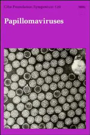 eBook - PDF
- David Evered, Sarah Clark, David Evered, Sarah Clark(Authors)
- 2008(Publication Date)
- Wiley(Publisher)
Saveria Campo raised the question of maternal transmission and of latency; an understanding of how these viruses are transmitted and of how long the latent phase can be before the clinical appearance of disease is necessary if we want to interpret studies designed to prevent or cure infections. Bill Pila- cinski described the expression of the BPV-1 capsid protein in bacteria and the use of fusion proteins as a vaccine in a limited trial. His studies show how little we know about the role of the immune system in controlling wart virus infec- tions and raise the question of whether vaccination is even a rational approach to attacking these infections. There are many different Papillomavirus-induced lesions in humans, and it now appears that certain Papillomaviruses are associ- ated with several types of human carcinoma: cutaneous carcinomas in epidermodysplasia verruciformis, anogenital and cervical carcinomas, and possibly oral carcinomas. Stefanie Jablonska and Gerard Orth have shown that, with the exception of one carcinoma in which they could not find papillo- mavirus DNA, a characteristic of carcinomas in epidermodysplasia verrucifor- mis is that the DNA remains extrachromosomal. This is in contrast to cervical carcinomas in which the HPV DNA appears to be integrated. Integration of the viral DNA occurs at a characteristic site with respect to the viral genome 246 HOWLEY around the E2/E1 junction. Lutz Gissmann’s results provide compelling evi- dence for a strong association between HPV types 16 and 18 and cervical, penile and vulvar carcinomas. Our understanding of the molecular biology of these two HPV types is progressing quickly. Bettie Steinberg has established that some of the Papillomaviruses of the genital tract are also present in the oropharynx and respiratory tract. eBook - PDF
- G.J. Galasso, C.A.B. Boucher, D.A. Cooper, D.A. Katzenstein(Authors)
- 2002(Publication Date)
- Elsevier Science(Publisher)
CHAPTER 11 PapillomavirusES RICHARD REICHMAN and MARGARET STANLEY Table of Contents General Introduction. ........................................................ 257 Clinical Diagnosis. .......................................... ............. 259 Genome Detection ...................................... Laboratory Diagnosis ..... ......................... 260 Serology ......... HPV testing In cervi .............. 261 When To Treat ......................... ........................... 262 Treatment Algorithms Cutaneous Warts ... .................... 262 Anogenital Warts.. Treatment Algorithms For Intra-epithelial Genital HPV Disease ...................... 264 HGSIL (CIN2 and 3) ....................................................... 264 .................... 265 Vulva1 intra-epithelial neoplasia (VIN) ......................................... 266 LGSIL (CIN 1). ... Immunobiology of HPV and prospects For immunotherapy of intra-epithelial genital HPV disease ................................................... Immunomodulators ................... 267 Special considerations For subgroups of patients . ...................... 269 Prophylaxis and vaccination strategies. ...................... References. ................................................................ 270 General Introduction Human Papillomaviruses (HPV) infect epithelial tissues of skin and mucous membranes, producing both benign and malignant neoplasms [ 11. Alternatively, infection may be asymptomatic, leading to a latent state and the possible development of disease after prolonged periods of time. HPV are members of the Papillomavirus genus of the family Papovaviridae. They are nonenveloped viruses, 50 to 55 nm in diameter, and have 257 Practical Guidelines in Antiviral Therapy Ed. by Charles A.B. Boucher and George J . Galusso. 257 - 218 0 2002 Elsevier Science. Printed in the Netherlands. 25 8 R. Reichman and M. Stanley icosahedral capsids containing a double-stranded, circular DNA genome. eBook - PDF
- Joseph Jordan, Albert Singer, Howard Jones, Mahmood Shafi, Joseph Jordan, Albert Singer, Howard Jones, Mahmood Shafi(Authors)
- 2009(Publication Date)
- Wiley-Blackwell(Publisher)
Given sufficient sensitivity and adequate tumour sampling, it is likely that virtually all cervical tumours contain HPV DNA (Walboomers and Meijer, 1997). Role of HPV HPVs are small, double-stranded DNA viruses of approxim-ately 55 nm with an icosahedral protein capsid containing 72 capsomers (Fig. 18.4). The genome is circular and contains 7500–8000 basepairs. Taxonomically, Papillomaviruses used to be a subfamily in the Papovaviridae family but are now grouped independently as a family, the Papillomaviridae, affecting both cutaneous and mucosal epithelia (Fig. 18.5). As infectious agents, they are highly specific to their hosts. Different HPVs are classified as types on the basis of DNA sequence homology in particular genes, specifically L1, which codes for the viral capsid, and E6 and E7, both of which have E5 HPV 16 L2 L1 E6 E7 E1 E4 E2 Fig. 18.4 Schematic of the human Papillomavirus 16 (HPV 16) genome, showing the arrangement of the major non-structural and capsid genes. The three circles correspond to the three reading frames in which a sense strand can be translated. There are no known gene products produced by the antisense strand. Viral protein early 4 (E4) is encoded by a messenger RNA transcript that includes the initial amino acids of the E1 gene. The region between late 1 (L1) and E6 is an important transcription regulatory region a the mRNAs encoding most non-structural (E6, E7, E1, E2, E4 and E5) and capsid (L1 and L2) genes originate in this region. Most Papillomavirus genomes resemble HPV 16 in general organisation. From Frazer (2004) with permission from Nature Publishing Group.
Index pages curate the most relevant extracts from our library of academic textbooks. They’ve been created using an in-house natural language model (NLM), each adding context and meaning to key research topics.